An Attention-based Weakly Supervised framework for Spitzoid Melanocytic Lesion Diagnosis in WSI
Publication
Metrics
AI Quick Summary
This paper proposes a novel weakly-supervised deep learning framework for diagnosing spitzoid melanocytic lesions using whole slide imaging (WSI). The model, based on inductive transfer learning, achieves high accuracy and assists pathologists by highlighting overlooked patterns in skin biopsies.
Paper Preview
Abstract
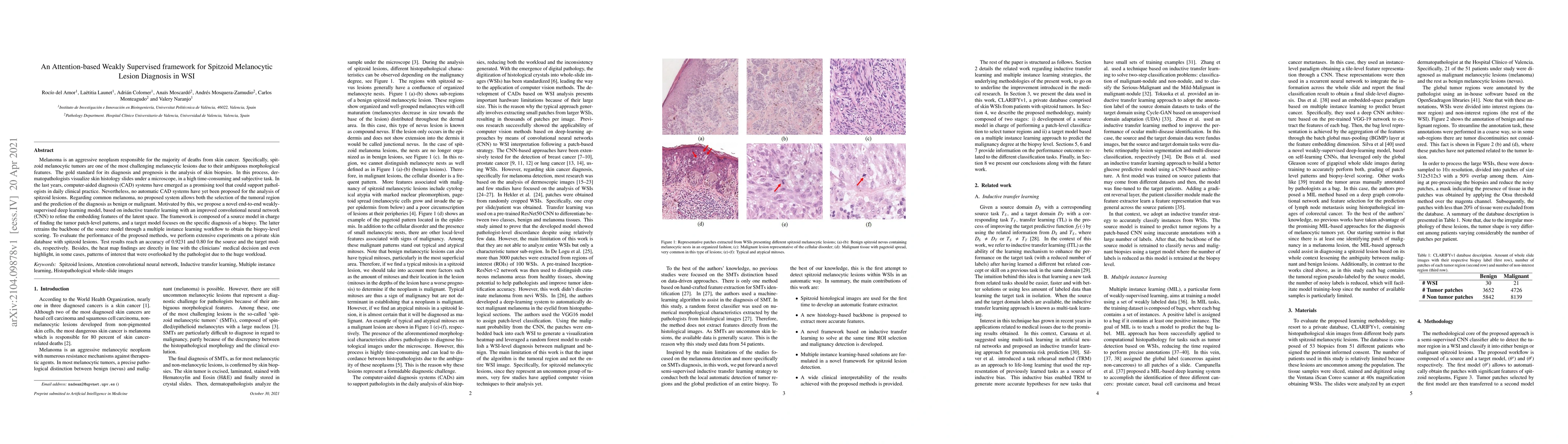
Melanoma is an aggressive neoplasm responsible for the majority of deaths from skin cancer. Specifically, spitzoid melanocytic tumors are one of the most challenging melanocytic lesions due to their ambiguous morphological features. The gold standard for its diagnosis and prognosis is the analysis of skin biopsies. In this process, dermatopathologists visualize skin histology slides under a microscope, in a high time-consuming and subjective task. In the last years, computer-aided diagnosis (CAD) systems have emerged as a promising tool that could support pathologists in daily clinical practice. Nevertheless, no automatic CAD systems have yet been proposed for the analysis of spitzoid lesions. Regarding common melanoma, no proposed system allows both the selection of the tumoral region and the prediction of the diagnosis as benign or malignant. Motivated by this, we propose a novel end-to-end weakly-supervised deep learning model, based on inductive transfer learning with an improved convolutional neural network (CNN) to refine the embedding features of the latent space. The framework is composed of a source model in charge of finding the tumor patch-level patterns, and a target model focuses on the specific diagnosis of a biopsy. The latter retrains the backbone of the source model through a multiple instance learning workflow to obtain the biopsy-level scoring. To evaluate the performance of the proposed methods, we perform extensive experiments on a private skin database with spitzoid lesions. Test results reach an accuracy of 0.9231 and 0.80 for the source and the target models, respectively. Besides, the heat map findings are directly in line with the clinicians' medical decision and even highlight, in some cases, patterns of interest that were overlooked by the pathologist due to the huge workload.
AI Key Findings
Get AI-generated insights about this paper's methodology, results, significance, and more — seven facets brought into focus.
Impact
Paper Details
Authors
PDF Preview
Key Terms
Citation Network
Current paper (gray), citations (green), references (blue)
Display is limited for performance on very large graphs.
Discussion 0