ArxivLens

Quick Summary:

This paper introduces an unsupervised method for segmenting overlapping cervical cell cytoplasm, addressing issues of poor contrast and cell overlap. It employs a modified Otsu method for nucleus detection and distance regularized level set evolution to delineate cytoplasm boundaries, achieving promising results on the ISBI 2015 challenge dataset.

Journal Reference: IEEE EMBS Conference on Biomedical Engineering and Sciences (IECBES), 2016
DOI: 10.1109/IECBES.2016.7843424
arXiv Id: 1702.05506
Comments: 4 pages, 4 figures, Biomedical Engineering and Sciences (IECBES), 2016 IEEE EMBS Conference on. IEEE, 2016
Date Published: 2018-07-16
Date Updated: 2018-07-16

Categories:

Key Terms (TF-IDF):

cell (0.327)
clumps (0.293)
segmentation (0.278)
overlapping (0.267)
boundaries (0.230)
nuclei (0.223)
accurate (0.175)
clump (0.173)
segments (0.161)
evolved (0.157)
reaches (0.157)
regularized (0.153)
poor (0.146)
cells (0.140)
unsupervised (0.140)
automated (0.139)
proposed approach (0.138)
nucleus (0.137)
results obtained (0.135)
paper presents (0.130)
License: http://arxiv.org/licenses/nonexclusive-distrib/1.0/

Summary:

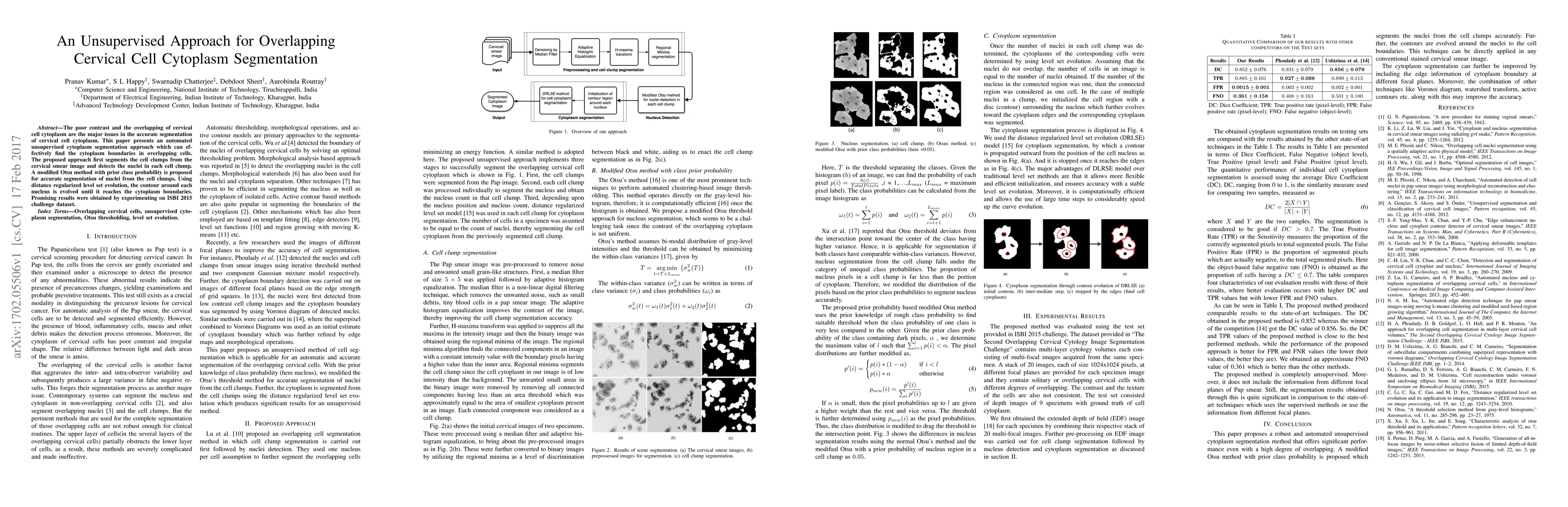
The poor contrast and the overlapping of cervical cell cytoplasm are the major issues in the accurate segmentation of cervical cell cytoplasm. This paper presents an automated unsupervised cytoplasm segmentation approach which can effectively find the cytoplasm boundaries in overlapping cells. The proposed approach first segments the cell clumps from the cervical smear image and detects the nuclei in each cell clump. A modified Otsu method with prior class probability is proposed for accurate segmentation of nuclei from the cell clumps. Using distance regularized level set evolution, the contour around each nucleus is evolved until it reaches the cytoplasm boundaries. Promising results were obtained by experimenting on ISBI 2015 challenge dataset.

Citation & Reference Network Visualization

This graph shows how this paper is connected to other research. The current paper is shown in dark gray, papers that cite it are in green, and papers it references are in blue. You can:

Current Paper
Papers citing this work (Citations)
Papers cited by this work (References)

• Hover over any node to see the paper title

• Click a node to view that paper's details

• Drag nodes to rearrange the graph

• Use the zoom controls (bottom right) to zoom in/out

Citations and References

Title Authors Year Actions
HVS-Unsup: Unsupervised cervical cell instance segmentation method based on human visual simulation
Xiaona Yang, Bo Ding, Jian Qin, Luyao Guo, Jing Zhao, Yongjun He
2024
View Paper
A Systematic Study of Challenges in Cervical Cytology Image Segmentation of Intelligence based Cervical Cancer Screening
P. M. Anu, C. Lakshmi
2023
View Paper
Cytology Image Analysis Techniques Toward Automation
Shyamali Mitra, N. Das, Soumyajyoti Dey, Sukanta Chakrabarty, M. Nasipuri, M. K. Naskar
2021
View Paper
Automated Segmentation of Overlapping Cells in Cervical Cytology Images Using Deep Learning
Ashwin Umadi, Kushal Nagarajan, Juthik Bangalore Venkatesha, Aashutosh Ganesh, Koshy George
2020
View Paper
Shape Mask Generator: Learning to Refine Shape Priors for Segmenting Overlapping Cervical Cytoplasms
Youyi Song, Lei Zhu, Baiying Lei, Bin Sheng, Q. Dou, J. Qin, K. Choi
2020
View Paper
Constrained Multi-shape Evolution for Overlapping Cytoplasm Segmentation
Youyi Song, Lei Zhu, Baiying Lei, Bin Sheng, Q. Dou, J. Qin, K. Choi
2020
View Paper
Cytology Image Analysis Techniques Towards Automation: Systematically Revisited
Shyamali Mitra, N. Das, Soumyajyoti Dey, Sukanta Chakrabarty, M. Nasipuri, M. K. Naskar
2020
View Paper
Computer-Assisted Screening for Cervical Cancer Using Digital Image Processing of Pap Smear Images
K. Win, Y. Kitjaidure, K. Hamamoto, Thet Myo Aung
2020
View Paper
Towards a Complete Pipeline for Segmenting Nuclei in Feulgen-Stained Images
Luiz Antonio Buschetto Macarini, A. V. Wangenheim, Felipe Perozzo Daltoé, A. S. Onofre, F. Onofre, M. Stemmer
2020
View Paper
Computational Nuclei Segmentation Methods in Digital Pathology: A Survey
Tomohiro Hayakawa, V. Prasath, H. Kawanaka, Bruce J. Aronow, Bruce J. Aronow, S. Tsuruoka
2019
View Paper
A Review of Computational Methods for Cervical Cells Segmentation and Abnormality Classification
T. Conceição, Cristiana Braga, Luís Rosado, M. Vasconcelos
2019
View Paper
Segmentation of Overlapping Cytoplasm in Cervical Smear Images via Adaptive Shape Priors Extracted From Contour Fragments
Youyi Song, Lei Zhu, J. Qin, Baiying Lei, Bin Sheng, K. Choi
2019
View Paper
A New Cervical Cytology Dataset for Nucleus Detection and Image Classification (Cervix93) and Methods for Cervical Nucleus Detection
H. A. Phoulady, P. Mouton
2018
View Paper
Multi-Pass Fast Watershed for Accurate Segmentation of Overlapping Cervical Cells
Afaf Tareef, Yang Song, Heng Huang, D. Feng, Mei Chen, Y. Wang, Weidong (Tom) Cai
2018
View Paper
Performance evaluation of extended depth of field microscopy in the presence of spherical aberration and noise
S. V. King, Shuai Yuan, C. Preza
2018
View Paper
Segmentation of overlapping cells obtained from pap smear test
H. Nisar, Lim Wai, Loh Siu Hong
2017
View Paper

Comments (0)

This website uses cookies to ensure you get the best experience. Learn more