Constrained Multi-shape Evolution for Overlapping Cytoplasm Segmentation
Publication
Metrics
AI Quick Summary
This paper introduces a constrained multi-shape evolution approach for segmenting overlapping cytoplasm in cervical smear images, addressing the challenge of intensity deficiency in overlapping regions. The method models both local and global shape priors to guide the segmentation process, ensuring the resulting shapes remain plausible and outperforms existing state-of-the-art methods.
Paper Preview
Abstract
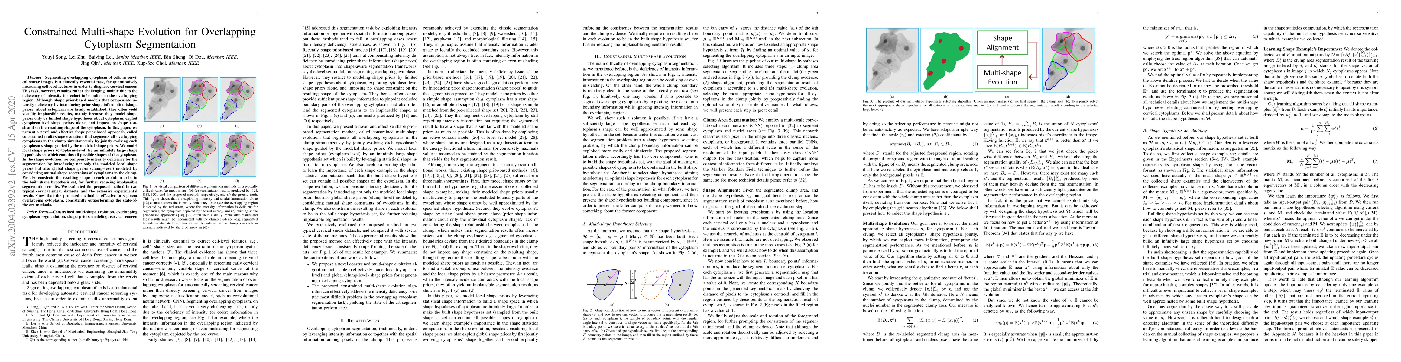
Segmenting overlapping cytoplasm of cells in cervical smear images is a clinically essential task, for quantitatively measuring cell-level features in order to diagnose cervical cancer. This task, however, remains rather challenging, mainly due to the deficiency of intensity (or color) information in the overlapping region. Although shape prior-based models that compensate intensity deficiency by introducing prior shape information (shape priors) about cytoplasm are firmly established, they often yield visually implausible results, mainly because they model shape priors only by limited shape hypotheses about cytoplasm, exploit cytoplasm-level shape priors alone, and impose no shape constraint on the resulting shape of the cytoplasm. In this paper, we present a novel and effective shape prior-based approach, called constrained multi-shape evolution, that segments all overlapping cytoplasms in the clump simultaneously by jointly evolving each cytoplasm's shape guided by the modeled shape priors. We model local shape priors (cytoplasm--level) by an infinitely large shape hypothesis set which contains all possible shapes of the cytoplasm. In the shape evolution, we compensate intensity deficiency for the segmentation by introducing not only the modeled local shape priors but also global shape priors (clump--level) modeled by considering mutual shape constraints of cytoplasms in the clump. We also constrain the resulting shape in each evolution to be in the built shape hypothesis set, for further reducing implausible segmentation results. We evaluated the proposed method in two typical cervical smear datasets, and the extensive experimental results show that the proposed method is effective to segment overlapping cytoplasm, consistently outperforming the state-of-the-art methods.
AI Key Findings
Get AI-generated insights about this paper's methodology, results, significance, and more — seven facets brought into focus.
Impact
Paper Details
Authors
PDF Preview
Key Terms
Citation Network
Current paper (gray), citations (green), references (blue)
Display is limited for performance on very large graphs.
Discussion 0