Quick Summary:

Researchers developed a new method for modeling image-derived input functions in dynamic PET scans, incorporating multiple organ segmentation to improve accuracy. This approach resulted in a 13.39% reduction in mean squared error for liver and 10.42% for lungs.

Summary

Accurate kinetic analysis of [$^{18}$F]FDG distribution in dynamic positron emission tomography (PET) requires anatomically constrained modelling of image-derived input functions (IDIFs). Traditionally, IDIFs are obtained from the aorta, neglecting anatomical variations and complex vascular contributions. This study proposes a multi-organ segmentation-based approach that integrates IDIFs from the aorta, portal vein, pulmonary artery, and ureters. Using high-resolution CT segmentations of the liver, lungs, kidneys, and bladder, we incorporate organ-specific blood supply sources to improve kinetic modelling. Our method was evaluated on dynamic [$^{18}$F]FDG PET data from nine patients, 
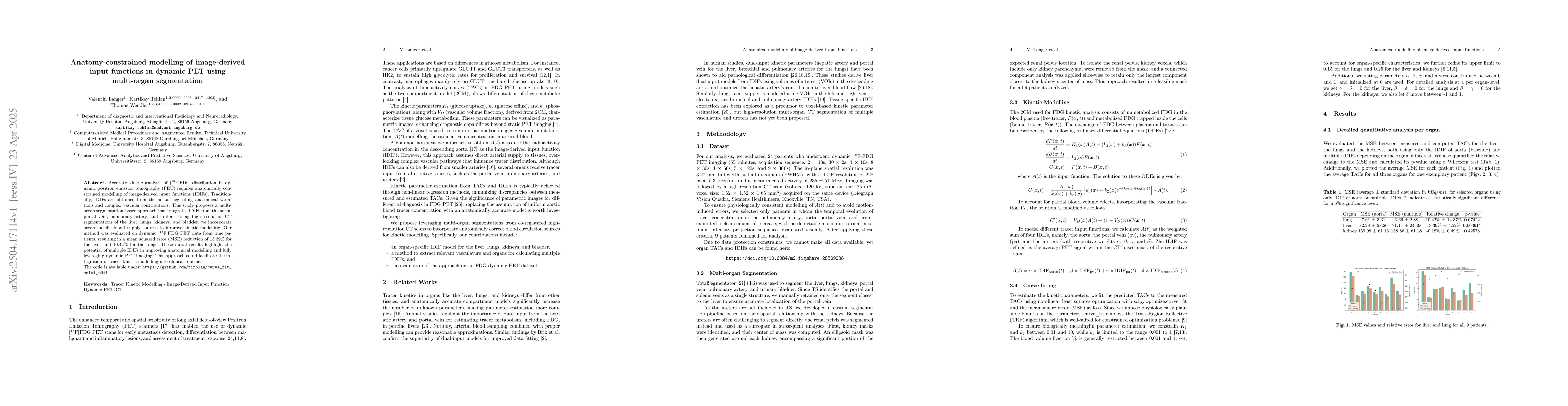
resulting in a mean squared error (MSE) reduction of $13.39\%$ for the liver and $10.42\%$ for the lungs. These initial results highlight the potential of multiple IDIFs in improving anatomical modelling and fully leveraging dynamic PET imaging. This approach could facilitate the integration of tracer kinetic modelling into clinical routine.

AI Key Findings

Generated Jun 09, 2025

Methodology

The study proposes a multi-organ segmentation-based approach for dynamic PET kinetic modeling, incorporating image-derived input functions (IDIFs) from the aorta, portal vein, pulmonary artery, and ureters using high-resolution CT segmentations of the liver, lungs, kidneys, and bladder.

Key Results

  • The method resulted in a mean squared error (MSE) reduction of 13.39% for the liver and 10.42% for the lungs when using multiple IDIFs compared to a single aortic IDIF.
  • The results highlight the potential of multiple IDIFs in improving anatomical modeling and leveraging dynamic PET imaging.
  • The approach showed a statistically significant improvement in modeling accuracy for the liver (p=0.00391) but not for the kidneys.

Significance

This research is important as it enhances the accuracy of tracer kinetic modeling in dynamic PET imaging, which is crucial for precise metabolic assessment in oncological applications.

Technical Contribution

The main technical contribution is the development of a method that integrates multiple anatomy-constrained IDIFs from various organ-specific blood supply sources to improve kinetic modeling in dynamic PET.

Novelty

This work is novel in its comprehensive approach to modeling tracer kinetics by considering multiple vascular input sources, moving beyond the traditional single aortic IDIF, and demonstrating its efficacy in improving modeling accuracy for organs like the liver.

Limitations

  • The study was limited by a small patient cohort, which might affect the generalizability of the results, particularly for the kidneys where improvements were minimal.
  • The approximation of ureter segmentation using the renal pelvis as a surrogate may not fully capture ureteral tracer kinetics.

Future Work

  • Future studies should explore more accurate ureter segmentation to enhance accuracy and better reflect renal excretion dynamics.
  • Further optimizations may be needed for other organs to fully leverage the multiple IDIF approach.

Paper Details

PDF Preview

Download PDF

Key Terms

No key terms available for this paper.

Citation Network

Current paper (gray), citations (green), references (blue)

Current Paper
Citations
References
Click to view

No citations found for this paper.

Comments (0)

This website uses cookies to ensure you get the best experience. Learn more