Dynamic FDG PET imaging study of n = 52 rats including 26 control
Wistar-Kyoto (WKY) rats and 26 experimental spontaneously hypertensive rats
(SHR) were performed using a Siemens microPET and Albira trimodal scanner
longitudinally at 1, 2, 3, 5, 9, 12 and 18 months of age. A 15-parameter dual
output model correcting for spill over contamination and partial volume effects
with peak fitting cost functions was developed for simultaneous estimation of
model corrected blood input function (MCIF) and kinetic rate constants for
dynamic FDG PET images of rat heart in vivo. Major drawbacks of this model are
its dependence on manual annotations for the Image Derived Input Function
(IDIF) and manual determination of crucial model parameters to compute MCIF. To
overcome these limitations, we performed semi-automated segmentation and then
formulated a Long-Short-Term Memory (LSTM) cell network to train and predict
MCIF in test data using a concatenation of IDIFs and myocardial inputs and
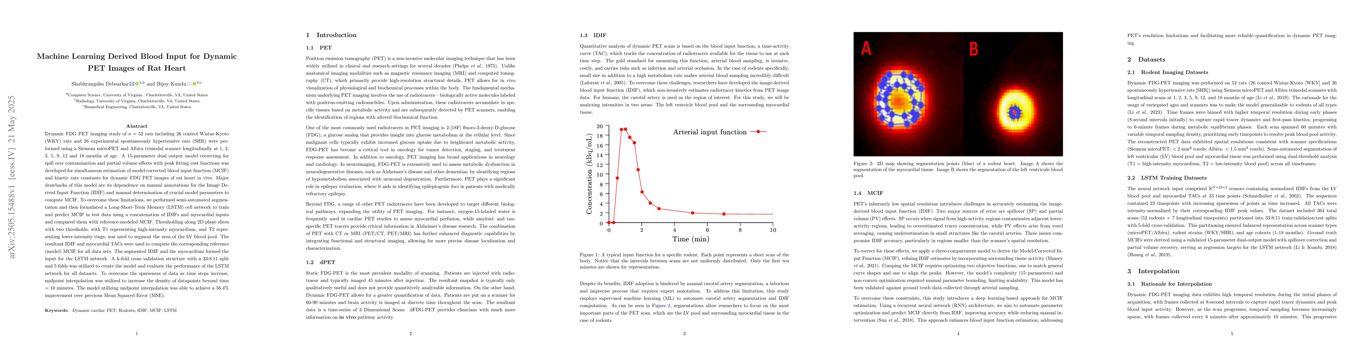
compared them with reference-modeled MCIF. Thresholding along 2D plane slices
with two thresholds, with T1 representing high-intensity myocardium, and T2
representing lower-intensity rings, was used to segment the area of the LV
blood pool. The resultant IDIF and myocardial TACs were used to compute the
corresponding reference (model) MCIF for all data sets. The segmented IDIF and
the myocardium formed the input for the LSTM network. A k-fold cross validation
structure with a 33:8:11 split and 5 folds was utilized to create the model and
evaluate the performance of the LSTM network for all datasets. To overcome the
sparseness of data as time steps increase, midpoint interpolation was utilized
to increase the density of datapoints beyond time = 10 minutes. The model
utilizing midpoint interpolation was able to achieve a 56.4% improvement over
previous Mean Squared Error (MSE).
Discussion 0