ArxivLens

Quick Summary:

This paper presents an approximate lesion localization method for dermoscopy images, utilizing an iterative algorithm to remove the black frame and an ensemble of thresholding algorithms to determine lesion location. The method was tested on 428 images, showing fast and accurate lesion localization compared to dermatologist-determined borders.

Journal Reference: Skin Research and Technology 15 (2009) 314-322
DOI: 10.1111/j.1600-0846.2009.00357.x
arXiv Id: 1009.1362
Date Published: 2010-09-08
Date Updated: 2010-09-08

Categories:

Key Terms (TF-IDF):

localization (0.282)
localization (0.281)
images (0.260)
images (0.250)
approximate (0.205)
approximate (0.196)
step (0.188)
determined (0.187)
step (0.184)
determined (0.182)
skin (0.175)
method (0.172)
demonstrate method (0.166)
method (0.166)
removed (0.165)
quantified (0.162)
modalities (0.161)
serves (0.161)
ground truth (0.158)
demonstrate method (0.157)
License: http://arxiv.org/licenses/nonexclusive-distrib/1.0/

Summary:

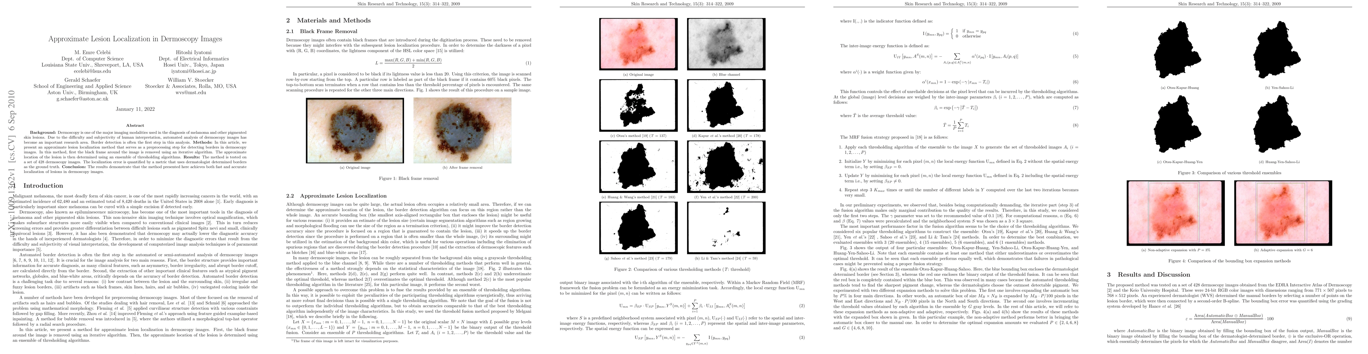
Background: Dermoscopy is one of the major imaging modalities used in the diagnosis of melanoma and other pigmented skin lesions. Due to the difficulty and subjectivity of human interpretation, automated analysis of dermoscopy images has become an important research area. Border detection is often the first step in this analysis. Methods: In this article, we present an approximate lesion localization method that serves as a preprocessing step for detecting borders in dermoscopy images. In this method, first the black frame around the image is removed using an iterative algorithm. The approximate location of the lesion is then determined using an ensemble of thresholding algorithms. Results: The method is tested on a set of 428 dermoscopy images. The localization error is quantified by a metric that uses dermatologist determined borders as the ground truth. Conclusion: The results demonstrate that the method presented here achieves both fast and accurate localization of lesions in dermoscopy images.

Citation & Reference Network Visualization

This graph shows how this paper is connected to other research. The current paper is shown in dark gray, papers that cite it are in green, and papers it references are in blue. You can:

Current Paper
Papers citing this work (Citations)
Papers cited by this work (References)

• Hover over any node to see the paper title

• Click a node to view that paper's details

• Drag nodes to rearrange the graph

• Use the zoom controls (bottom right) to zoom in/out

Citations and References

Title Authors Year Actions
Artificial intelligence in melanoma: A systematic review
Shu Zhang, Yuanzhuo Wang, Qingyue Zheng, Jiarui Li, Jiuzuo Huang, Xiao Long
2022
View Paper
A Survey on Deep Learning for Skin Lesion Segmentation
Z. Mirikharaji, Catarina Barata, Kumar Abhishek, Alceu Bissoto, S. Avila, Eduardo Valle, M. Celebi, G. Hamarneh
2022
View Paper
Deep Learning Approaches Towards Skin Lesion Segmentation and Classification from Dermoscopic Images - A Review.
R. Baig, Maryam Bibi, Anmol Hamid, Sumaira Kausar, Shahzad Khalid
2020
View Paper
Accurate Segmentation of Dermoscopic Images based on Local Binary Pattern Clustering
Pedro M. M. Pereira, R. Fonseca-Pinto, Rui Pedro Paiva, Luís M. N. Tavora, P. Assunção, S. Faria
2019
View Paper
Sampling with level set for pigmented skin lesion segmentation
Tiejun Yang, Yaowen Chen, Jiewei Lu, Zhun Fan
2019
View Paper
Region of Interest Detection in Dermoscopic Images for Natural Data-augmentation
M. Goyal, Moi Hoon Yap
2018
View Paper
Recent advancement in the early detection of melanoma using computerized tools: An image analysis perspective
Nivedita Singh, S. Gupta
2018
View Paper
A Review of Prevalent Methods for Automatic Skin Lesion Diagnosis
D. Okuboyejo, O. Olugbara
2018
View Paper
Computational methods for pigmented skin lesion classification in images: review and future trends
R. B. Oliveira, J. Papa, A. S. Pereira, J. Tavares
2018
View Paper
Segmentation of skin lesions using an improved FLICM method
S. Kanaani, M. Helfroush, H. Danyali, M. A. Kazemi
2017
View Paper
Pattern Recognition in Pigmented Skin Lesion Images using Ensemble Methods
R. B. Oliveira
2017
View Paper
An Overview of Melanoma Detection in Dermoscopy Images Using Image Processing and Machine Learning
N. Mishra, M. E. Celebi
2016
View Paper
A New fuzzy level set method for lesion border detection in dermoscopy images
S. V. Shojaedini
2015
View Paper
A State-of-the-Art Survey on Lesion Border Detection in Dermoscopy Images
M. E. Celebi, Q. Wen, H. Iyatomi, Kouhei Shimizu, Huiyu Zhou, G. Schaefer
2015
View Paper
Variability in human and automated segmentation of dermoscopy images of melanocytic skin lesions
Bogo Federica, Peruch Francesco, A. B. Fortina, P. Enoch
2015
View Paper
Automatic change detection in multiple pigmented skin lesions
K. Korotkov
2014
View Paper
Image thresholding based on semivariance
M. Beauchemin
2013
View Paper
Computerized analysis of pigmented skin lesions: A review
K. Korotkov, Rafael García
2012
View Paper
Fast density-based lesion detection in dermoscopy images
Mutlu Mete, S. Kockara, K. Aydin
2011
View Paper
Analysis of density based and fuzzy c-means clustering methods on lesion border extraction in dermoscopy images
S. Kockara, Mutlu Mete, Bernard Chen, K. Aydin
2010
View Paper
Contrast enhancement in dermoscopy images by maximizing a histogram bimodality measure
M. E. Celebi, H. Iyatomi, G. Schaefer
2009
View Paper
Localization of Lesions in Dermoscopy Images Using Ensembles of Thresholding Methods
M. E. Celebi, H. Iyatomi, G. Schaefer, W. Stoecker
2009
View Paper
Skin Lesion Detection by Android Camera based on SSD- Mo-bilenet and TensorFlow Object Detection API
A. Taqi, Fadwa Al-Azzo, AhmedM.El Awad, M. Milanova
2019
View Paper
Techniques and algorithms for computer aided diagnosis of pigmented skin lesions - A review
Sameena Pathan, K. G. Prabhu, P. Siddalingaswamy
2018
View Paper
Computing Equivalent Affinity Classes in a Fuzzy Connectedness Framework
G. Gulyás, J. Dombi
2014
View Paper
A scheme of extracting age-related wrinkle feature and skin age based on dermoscopic images
Young-Hwan Choi, Eenjun Hwang
2010
View Paper

Comments (0)

This website uses cookies to ensure you get the best experience. Learn more