Lesion Border Detection in Dermoscopy Images
Publication
Metrics
AI Quick Summary
This paper reviews recent methods for detecting lesion borders in dermoscopy images, highlighting computational challenges and evaluation issues. It concludes that incorporating domain knowledge into automated methods could improve performance, especially for diverse diagnostic sets.
Paper Preview
Abstract
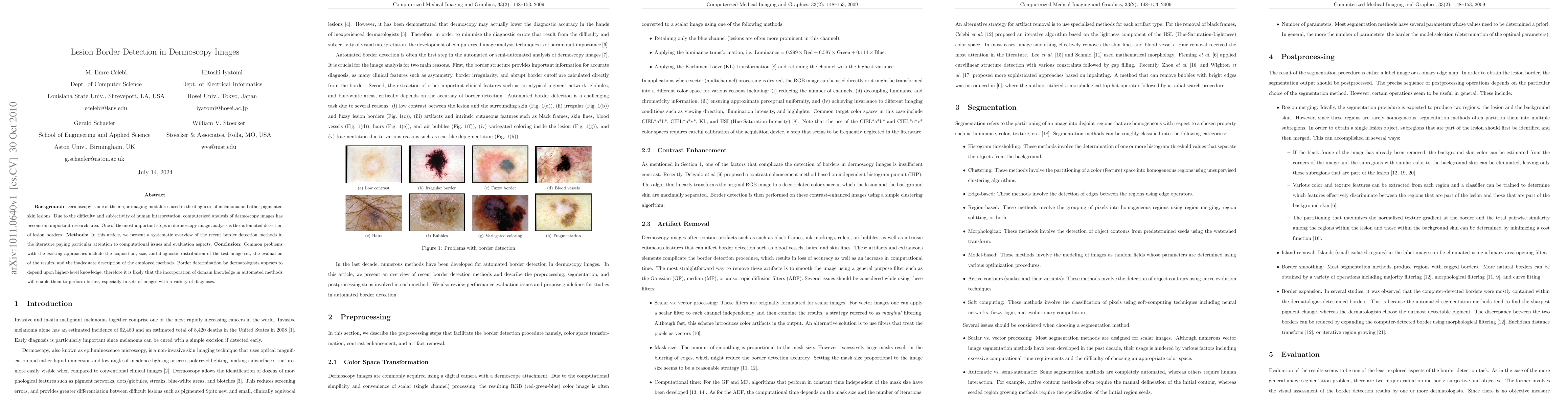
Background: Dermoscopy is one of the major imaging modalities used in the diagnosis of melanoma and other pigmented skin lesions. Due to the difficulty and subjectivity of human interpretation, computerized analysis of dermoscopy images has become an important research area. One of the most important steps in dermoscopy image analysis is the automated detection of lesion borders. Methods: In this article, we present a systematic overview of the recent border detection methods in the literature paying particular attention to computational issues and evaluation aspects. Conclusion: Common problems with the existing approaches include the acquisition, size, and diagnostic distribution of the test image set, the evaluation of the results, and the inadequate description of the employed methods. Border determination by dermatologists appears to depend upon higher-level knowledge, therefore it is likely that the incorporation of domain knowledge in automated methods will enable them to perform better, especially in sets of images with a variety of diagnoses.
AI Key Findings
Get AI-generated insights about this paper's methodology, results, significance, and more — seven facets brought into focus.
Impact
Paper Details
PDF Preview
Key Terms
Citation Network
Current paper (gray), citations (green), references (blue)
Display is limited for performance on very large graphs.
Discussion 0