AugHover-Net: Augmenting Hover-net for Nucleus Segmentation and Classification
Publication
Metrics
AI Quick Summary
The paper proposes an augmented version of Hover-Net for nucleus segmentation and classification in digital pathology, addressing challenges like dataset annotation, nucleus clustering, class imbalance, and inter-class similarity. The method was developed for the CoNIC challenge, aiming for efficient segmentation and classification of six nucleus types using the Lizard dataset.
Paper Preview
Abstract
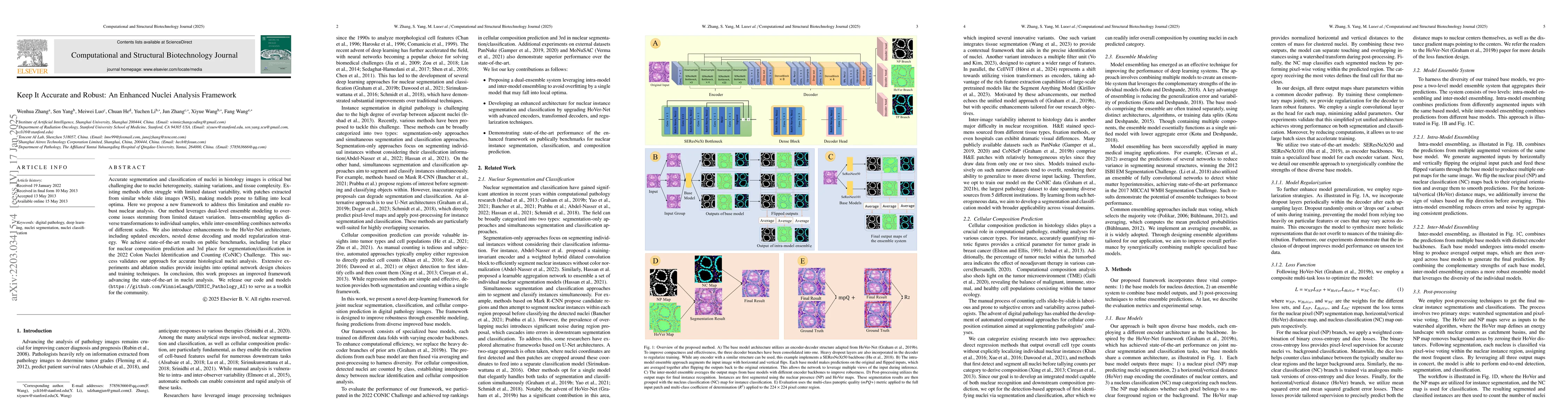
Nuclei segmentation and classification have been a challenge in digital pathology due to the specific domain characteristics. First, annotating a large-scale dataset is quite consuming. It requires specific domain knowledge and large efforts. Second, some nuclei are clustered together and hard to segment from each other. Third, the classes are often extremely unbalanced. As in Lizard, the number of epithelial nuclei is around 67 times larger than the number of eosinophil nuclei. Fourth, the nuclei often exhibit high inter-class similarity and intra-class variability. Connective nuclei may look very different from each other while some of them share a similar shape with the epithelial ones. Last but not least, pathological patches may have very different color distributions among different datasets. Thus, a large-scale generally annotated dataset and a specially-designed algorithm are needed to solve this problem. The CoNIC challenge aims to promote the automatic segmentation and classification task and requires researchers to develop algorithms that perform segmentation, classification, and counting of 6 different types of nuclei with the large-scale annotated dataset: Lizard. Due to the 60-minute time limit, the algorithm has to be simple and quick. In this paper, we briefly describe the final method we used in the CoNIC challenge. Our algorithm is based on Hover-Net and we added several modifications to it to improve its performance.
AI Key Findings
Get AI-generated insights about this paper's methodology, results, significance, and more — seven facets brought into focus.
Impact
Paper Details
PDF Preview
Key Terms
Citation Network
Current paper (gray), citations (green), references (blue)
Display is limited for performance on very large graphs.
Related Papers
No references found for this paper.
Discussion 0