ArxivLens

Quick Summary:

This paper presents a Color Space-based HoVer-Net model for nuclei instance segmentation and classification, utilizing ConvNeXt encoder, multi-channel color space, Unified Focal loss, and Sharpness-Aware Minimization to address segmentation challenges, achieving a 12.489% improvement over the current SOTA on the CoNiC Challenge 2022 test set.

arXiv Id: 2203.01940
Comments: 3 pages, 1 figure, and 1 table
Date Published: 2022-03-07
Date Updated: 2022-03-07

Categories:

Key Terms (TF-IDF):

nuclei (0.249)
nuclei (0.247)
imbalance (0.247)
net (0.213)
segmentation (0.212)
net (0.209)
segmentation (0.201)
differences (0.188)
differences (0.180)
used (0.152)
used (0.151)
sota (0.148)
based approach (0.146)
current state (0.142)
distinguishing (0.142)
extracting (0.141)
aid (0.141)
current state (0.140)
transformers (0.140)
suffers (0.140)
License: http://creativecommons.org/licenses/by-nc-sa/4.0/

Summary:

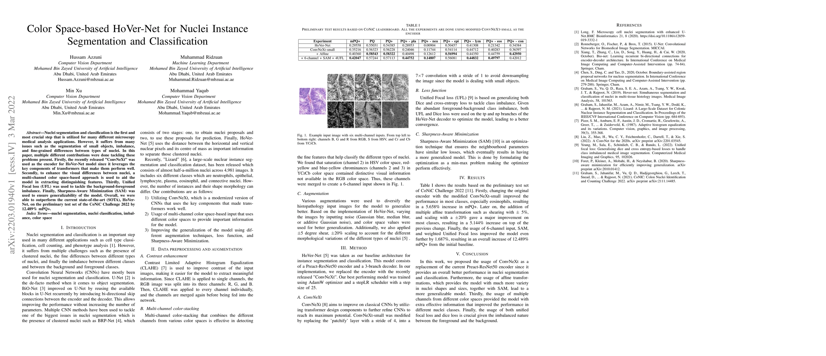
Nuclei segmentation and classification is the first and most crucial step that is utilized for many different microscopy medical analysis applications. However, it suffers from many issues such as the segmentation of small objects, imbalance, and fine-grained differences between types of nuclei. In this paper, multiple different contributions were done tackling these problems present. Firstly, the recently released "ConvNeXt" was used as the encoder for HoVer-Net model since it leverages the key components of transformers that make them perform well. Secondly, to enhance the visual differences between nuclei, a multi-channel color space-based approach is used to aid the model in extracting distinguishing features. Thirdly, Unified Focal loss (UFL) was used to tackle the background-foreground imbalance. Finally, Sharpness-Aware Minimization (SAM) was used to ensure generalizability of the model. Overall, we were able to outperform the current state-of-the-art (SOTA), HoVer-Net, on the preliminary test set of the CoNiC Challenge 2022 by 12.489% mPQ+.

Citation & Reference Network Visualization

This graph shows how this paper is connected to other research. The current paper is shown in dark gray, papers that cite it are in green, and papers it references are in blue. You can:

Current Paper
Papers citing this work (Citations)
Papers cited by this work (References)

• Hover over any node to see the paper title

• Click a node to view that paper's details

• Drag nodes to rearrange the graph

• Use the zoom controls (bottom right) to zoom in/out

Citations and References

Title Authors Year Actions
A Survey on Cell Nuclei Instance Segmentation and Classification: Leveraging Context and Attention
João D. Nunes, D. Montezuma, Domingos Oliveira, Tania Pereira, Jaime S. Cardoso
2024
View Paper
Counteracting Data Bias and Class Imbalance—Towards a Useful and Reliable Retinal Disease Recognition System
Adam R. Chłopowiec, Konrad Karanowski, Tomasz Skrzypczak, Mateusz Grzesiuk, Adrian B. Chłopowiec, M. Tabakov
2023
View Paper
An improved Hover-net for nuclear segmentation and classification in histopathology images
Ji Wang, Lulu Qin, Dan Chen, Juan Wang, Bo Han, Zexuan Zhu, Guangdong Qiao
2023
View Paper
Nuclear Segmentation and Classification: On Color & Compression Generalization
Q. Vu, R. Jewsbury, S. Graham, M. Jahanifar, S. Raza, F. Minhas, A. Bhalerao, N. Rajpoot
2023
View Paper
Transformers in Medical Image Analysis: A Review
Kelei He, Chen Gan, Zhuoyuan Li, I. Rekik, Zihao Yin, Wen Ji, Yang Gao, Qian Wang, Junfeng Zhang, D. Shen
2022
View Paper
Transformers in medical image analysis
Kelei He, Chen Gan, Zhuoyuan Li, I. Rekik, Zihao Yin, Wen Ji, Yang Gao, Qian Wang, Junfeng Zhang, Dinggang Shen
View Paper

Comments (0)

This website uses cookies to ensure you get the best experience. Learn more