Quick Answers
What methodology did the authors use?
The study compared five encoder-decoder architectures using 913 frames from 200 cases in the IARC Cervical Image Dataset, annotated by gynaecologists. Performance was evaluated using IoU, DICE, detection rate, and distance metrics with ten-fold cross-validation. More in Methodology →
What are the key results?
EndoViT/DPT achieved the highest DICE score (0.50 ± 0.31) and detection rate (0.87 ± 0.33) compared to other models. — External validation with phantom data showed robust segmentation under variable conditions at 21.5 FPS, supporting real-time feasibility. More in Key Results →
Why is this work significant?
This research addresses barriers to cervical cancer screening by enabling speculum-free, camera-guided devices with automated visual guidance, which could improve access in low-resource settings and support non-expert use. More in Significance →
What are the main limitations?
Performance metrics show variability (e.g., DICE ± 0.31), indicating potential inconsistency in segmentation results. — The study was limited to a specific dataset and may not generalize to all clinical scenarios. More in Limitations →
Abstract
Cervical cancer is highly preventable, yet persistent barriers to screening limit progress toward elimination goals. Speculum-free devices that integrate imaging and sampling could improve access, particularly in low-resource settings, but require reliable visual guidance. This study evaluates deep learning methods for real-time segmentation of the cervical os in transvaginal 
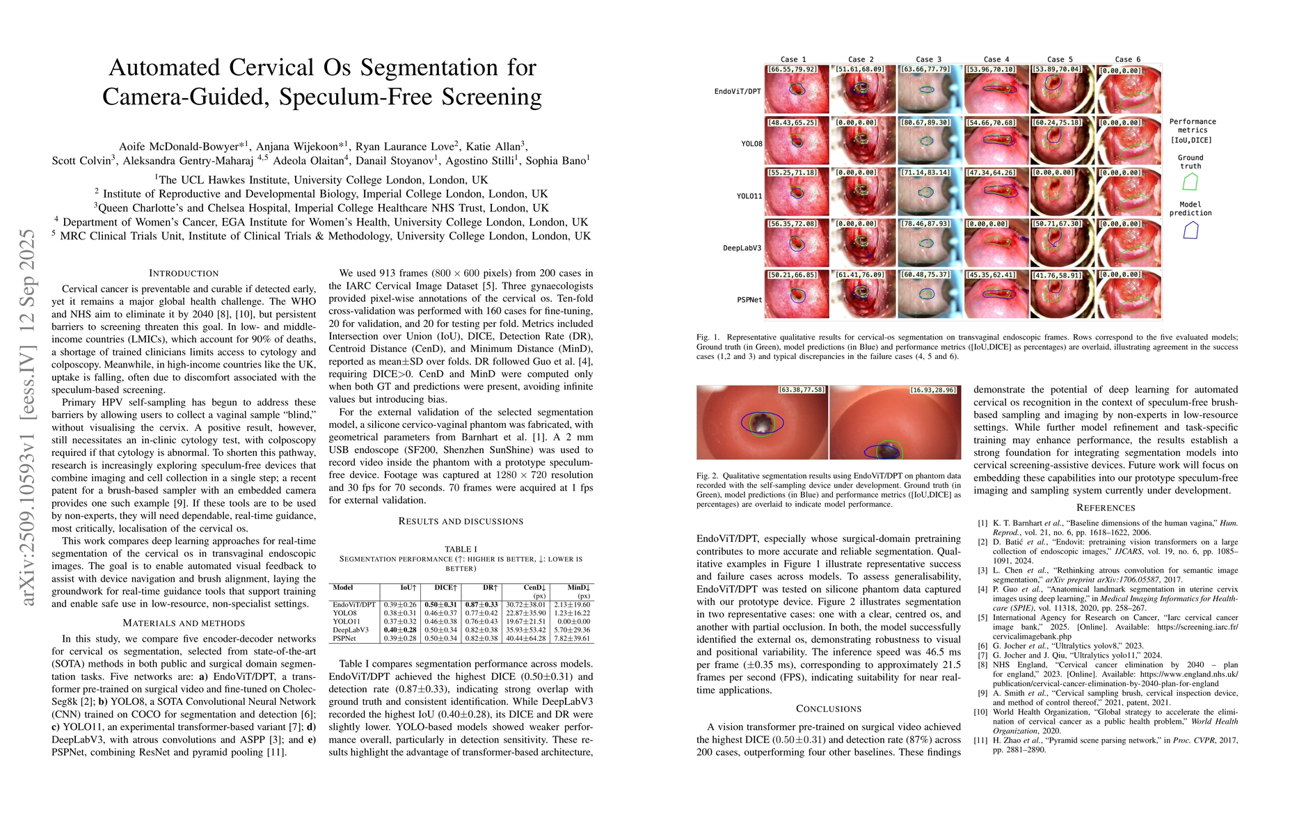
endoscopic images. Five encoder-decoder architectures were compared using 913 frames from 200 cases in the IARC Cervical Image Dataset, annotated by gynaecologists. Performance was assessed using IoU, DICE, detection rate, and distance metrics with ten-fold cross-validation. EndoViT/DPT, a vision transformer pre-trained on surgical video, achieved the highest DICE (0.50 \pm 0.31) and detection rate (0.87 \pm 0.33), outperforming CNN-based approaches. External validation with phantom data demonstrated robust segmentation under variable conditions at 21.5 FPS, supporting real-time feasibility. These results establish a foundation for integrating automated os recognition into speculum-free cervical screening devices to support non-expert use in both high- and low-resource contexts.
AI Key Findings
Generated Oct 18, 2025
Methodology — What approach did the authors take?
The study compared five encoder-decoder architectures using 913 frames from 200 cases in the IARC Cervical Image Dataset, annotated by gynaecologists. Performance was evaluated using IoU, DICE, detection rate, and distance metrics with ten-fold cross-validation.
Key Results — What are the main findings?
- EndoViT/DPT achieved the highest DICE score (0.50 ± 0.31) and detection rate (0.87 ± 0.33) compared to other models.
- External validation with phantom data showed robust segmentation under variable conditions at 21.5 FPS, supporting real-time feasibility.
- Transformer-based models outperformed CNN-based approaches in segmentation accuracy and detection consistency.
Significance — Why does this research matter?
This research addresses barriers to cervical cancer screening by enabling speculum-free, camera-guided devices with automated visual guidance, which could improve access in low-resource settings and support non-expert use.
Technical Contribution — What is the technical contribution?
Development and evaluation of a vision transformer pre-trained on surgical video for real-time cervical os segmentation in transvaginal endoscopic images.
Novelty — What is new about this work?
The use of a vision transformer pre-trained on surgical video (EndoViT/DPT) for cervical os segmentation, which outperforms traditional CNN-based approaches in both accuracy and real-time performance.
Limitations — What are the limitations of this study?
- Performance metrics show variability (e.g., DICE ± 0.31), indicating potential inconsistency in segmentation results.
- The study was limited to a specific dataset and may not generalize to all clinical scenarios.
Future Work — What did the authors propose for future work?
- Integrate segmentation models into prototype speculum-free imaging and sampling systems.
- Further refine models for task-specific training and improve performance under challenging conditions.
Paper Details
How to Cite This Paper
@article{olaitan2025automated,
title = {Automated Cervical Os Segmentation for Camera-Guided, Speculum-Free
Screening},
author = {Olaitan, Adeola and Stilli, Agostino and Gentry-Maharaj, Aleksandra and others},
year = {2025},
eprint = {2509.10593},
archivePrefix = {arXiv},
primaryClass = {eess.IV},
}
Olaitan, A., Stilli, A., Gentry-Maharaj, A., Wijekoon, A., McDonald-Bowyer, A., Stoyanov, D., Allan, K., Love, R., Colvin, S., & Bano, S. (2025). Automated Cervical Os Segmentation for Camera-Guided, Speculum-Free
Screening. arXiv. https://arxiv.org/abs/2509.10593
Olaitan, Adeola, et al. "Automated Cervical Os Segmentation for Camera-Guided, Speculum-Free
Screening." arXiv, 2025, arxiv.org/abs/2509.10593.
PDF Preview
Similar Papers
Found 4 papersAI Guided Early Screening of Cervical Cancer
Dharanidharan S I, Suhitha Renuka S V, Ajishi Singh et al.
A Novel, Versatile Speculum-free Callascope for Clinical Examination and Self-Visualization of the Cervix
Asiedu, M., Agudogo, J. S., Dotson, M. E. et al.
Automated CT Lung Cancer Screening Workflow using 3D Camera
Brian Teixeira, Vivek Singh, Birgi Tamersoy et al.
Segmentation Guided Sparse Transformer for Under-Display Camera Image Restoration
Jingyun Xue, Tao Wang, Jun Wang et al.
Comments (0)