Automated high-resolution backscattered-electron imaging at macroscopic scale
Publication
Metrics
AI Quick Summary
This research proposes a novel method to generate high-resolution SEM images that capture macroscopic dimensions up to centimeters while preserving submicron details. The approach combines SEM imaging, video stitching, and a denoising model, demonstrating its application on an AlCoCrFeNi2.1 eutectic high entropy alloy, revealing microstructure correlations with hardness.
Paper Preview
Abstract
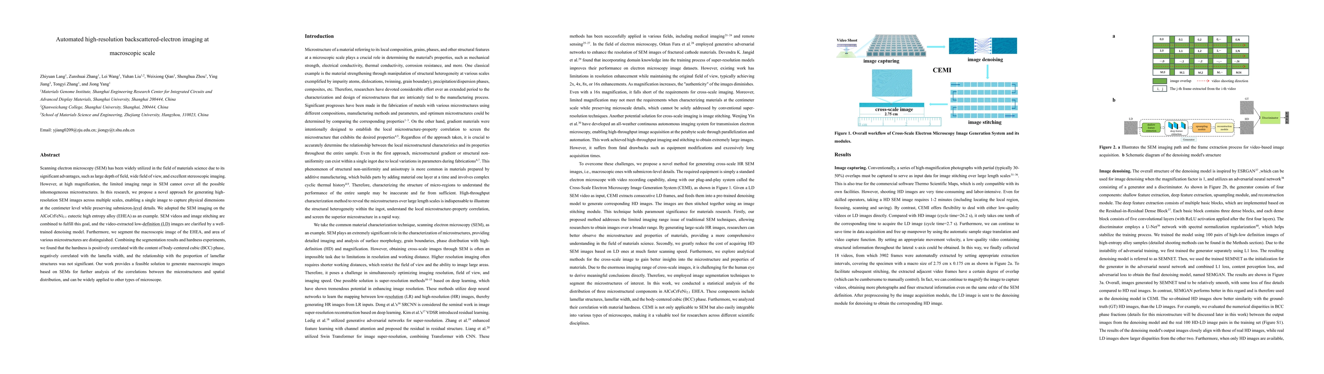
Scanning electron microscopy (SEM) has been widely utilized in the field of materials science due to its significant advantages, such as large depth of field, wide field of view, and excellent stereoscopic imaging. However, at high magnification, the limited imaging range in SEM cannot cover all the possible inhomogeneous microstructures. In this research, we propose a novel approach for generating high-resolution SEM images across multiple scales, enabling a single image to capture physical dimensions at the centimeter level while preserving submicron-level details. We adopted the SEM imaging on the AlCoCrFeNi2.1 eutectic high entropy alloy (EHEA) as an example. SEM videos and image stitching are combined to fulfill this goal, and the video-extracted low-definition (LD) images are clarified by a well-trained denoising model. Furthermore, we segment the macroscopic image of the EHEA, and area of various microstructures are distinguished. Combining the segmentation results and hardness experiments, we found that the hardness is positively correlated with the content of body-centered cubic (BCC) phase, negatively correlated with the lamella width, and the relationship with the proportion of lamellar structures was not significant. Our work provides a feasible solution to generate macroscopic images based on SEMs for further analysis of the correlations between the microstructures and spatial distribution, and can be widely applied to other types of microscope.
AI Key Findings — Failed
Key findings generation failed. Failed to start generation process
Impact
Paper Details
Authors
PDF Preview
Citation Network
Current paper (gray), citations (green), references (blue)
Display is limited for performance on very large graphs.
Discussion 0