01
MethodologyHow they did it
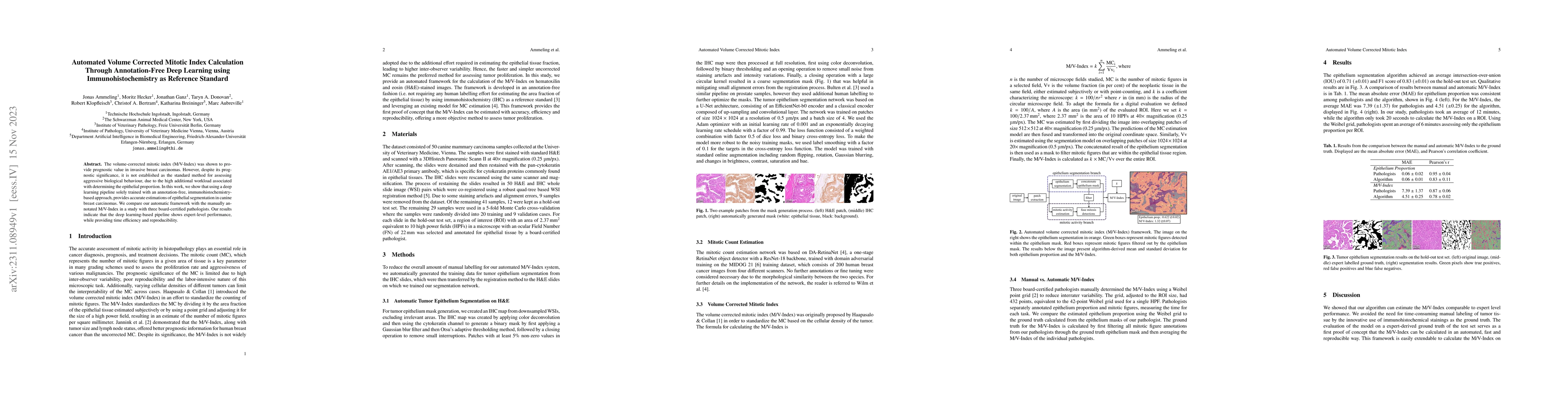
The research presents an automated framework for calculating the volume-corrected mitotic index (M/V-Index) on H&E-stained images using an annotation-free, immunohistochemistry (IHC)-based approach. The framework consists of two networks: one for tumor epithelium segmentation and another for mitotic count estimation. The epithelium segmentation network is based on U-Net with an EfficientNet-b0 encoder, while the mitotic count estimation network is based on DA-RetinaNet with a ResNet-18 backbone.
Discussion 0