Academic Profile
Statistics
Similar Authors
Papers on arXiv
Acquiring annotations for whole slide images (WSIs)-based deep learning tasks, such as creating tissue segmentation masks or detecting mitotic figures, is a laborious process due to the extensive imag...
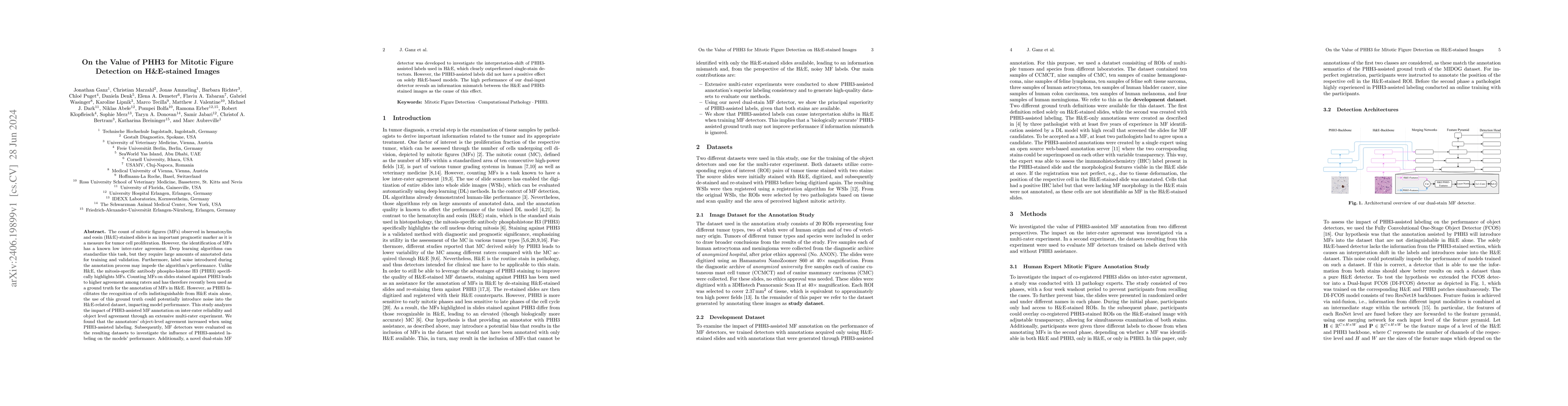
The count of mitotic figures (MFs) observed in hematoxylin and eosin (H&E)-stained slides is an important prognostic marker as it is a measure for tumor cell proliferation. However, the identification...
Background: This research aims to improve glioblastoma survival prediction by integrating MR images, clinical and molecular-pathologic data in a transformer-based deep learning model, addressing dat...
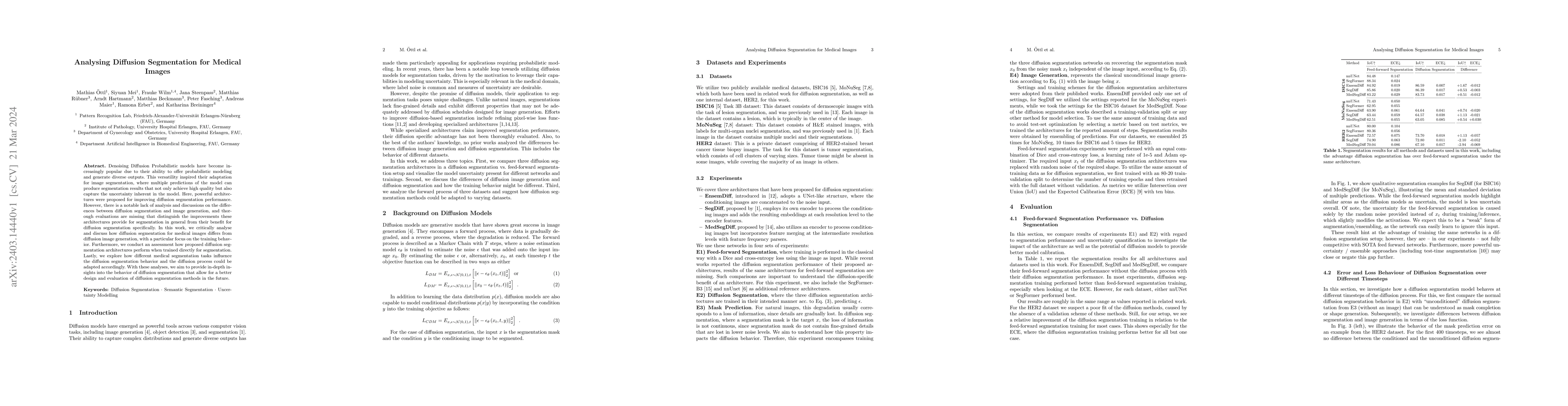
Denoising Diffusion Probabilistic models have become increasingly popular due to their ability to offer probabilistic modeling and generate diverse outputs. This versatility inspired their adaptatio...
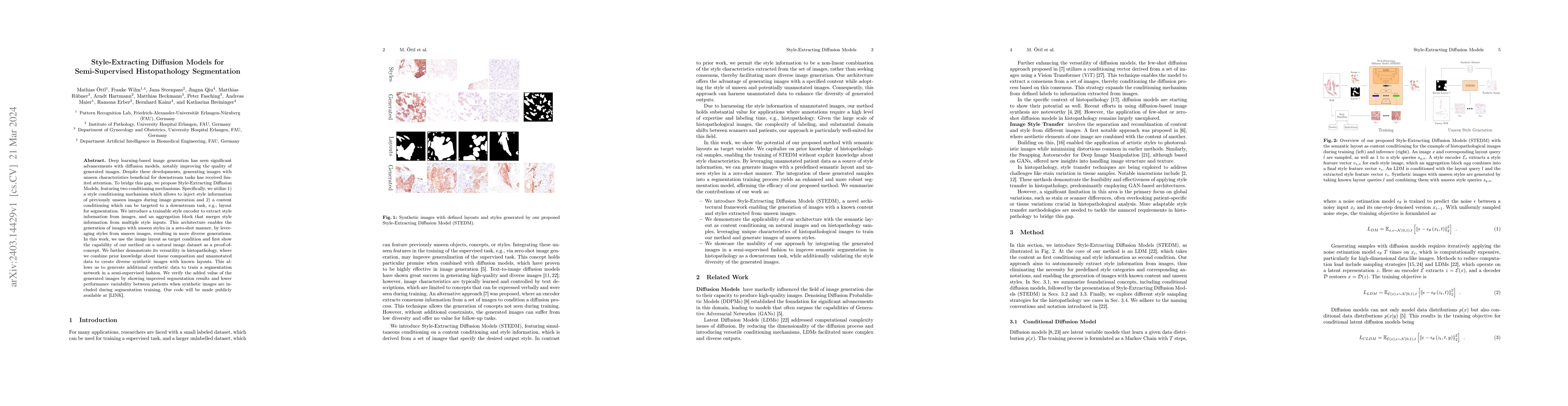
Deep learning-based image generation has seen significant advancements with diffusion models, notably improving the quality of generated images. Despite these developments, generating images with un...
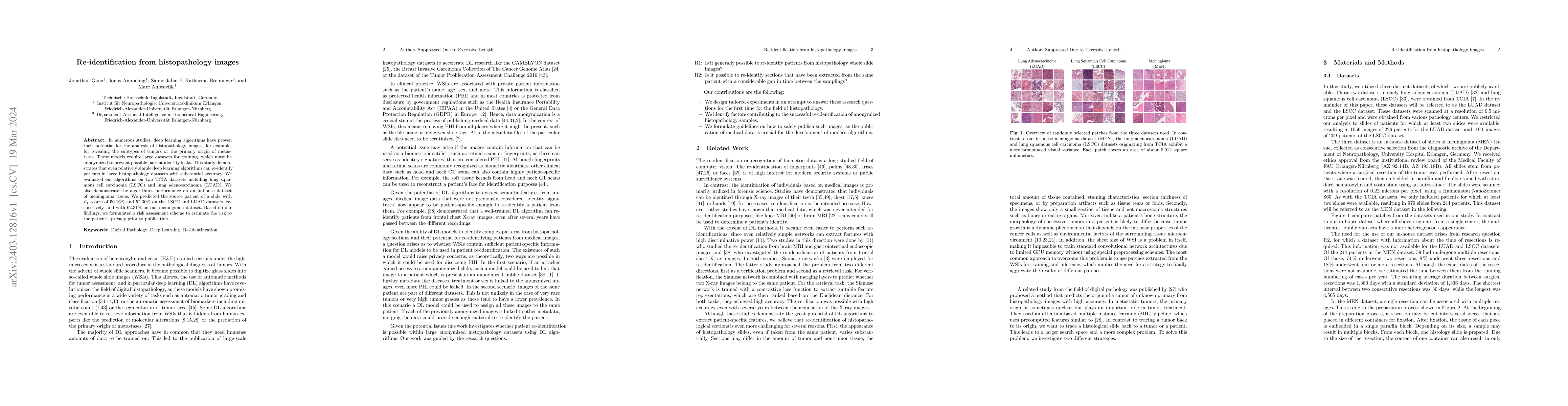
In numerous studies, deep learning algorithms have proven their potential for the analysis of histopathology images, for example, for revealing the subtypes of tumors or the primary origin of metast...
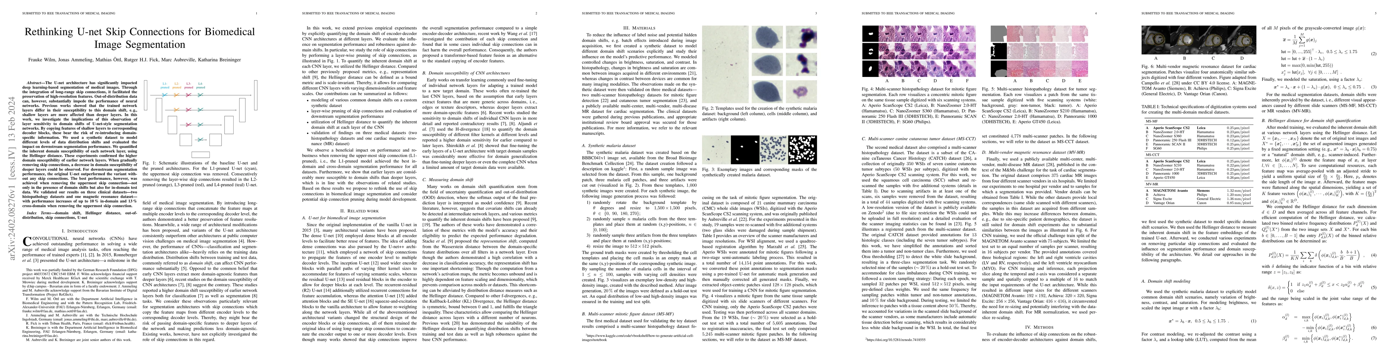
The U-net architecture has significantly impacted deep learning-based segmentation of medical images. Through the integration of long-range skip connections, it facilitated the preservation of high-...
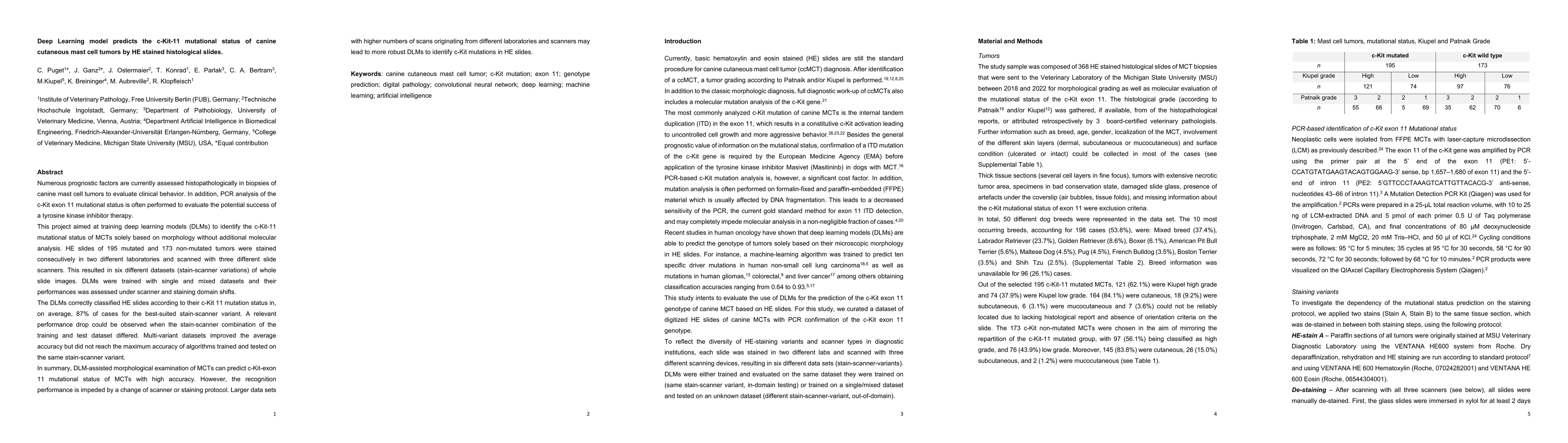
Numerous prognostic factors are currently assessed histopathologically in biopsies of canine mast cell tumors to evaluate clinical behavior. In addition, PCR analysis of the c-Kit exon 11 mutational...
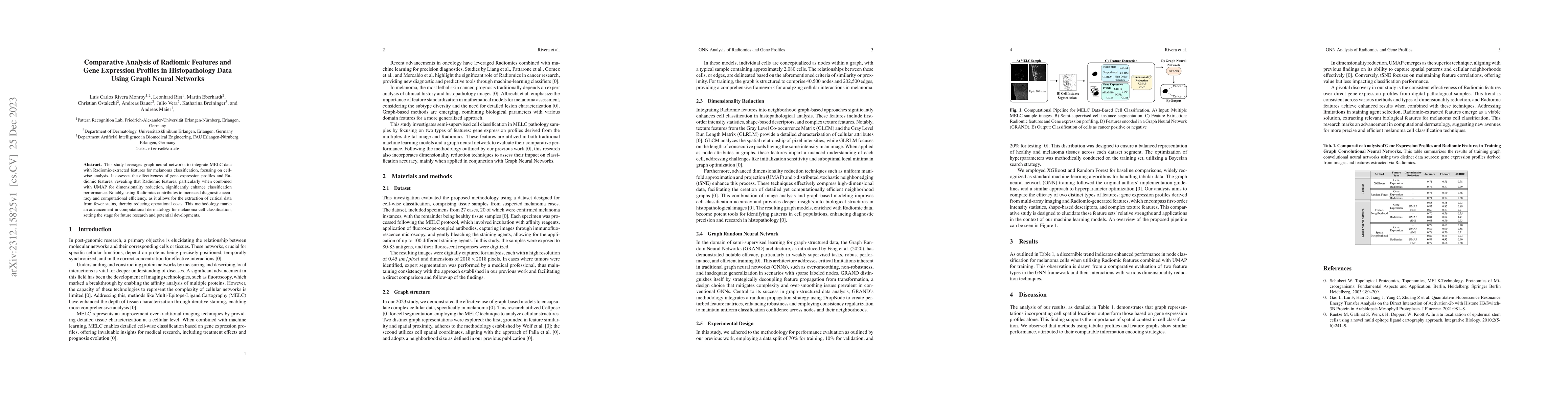
This study leverages graph neural networks to integrate MELC data with Radiomic-extracted features for melanoma classification, focusing on cell-wise analysis. It assesses the effectiveness of gene ...
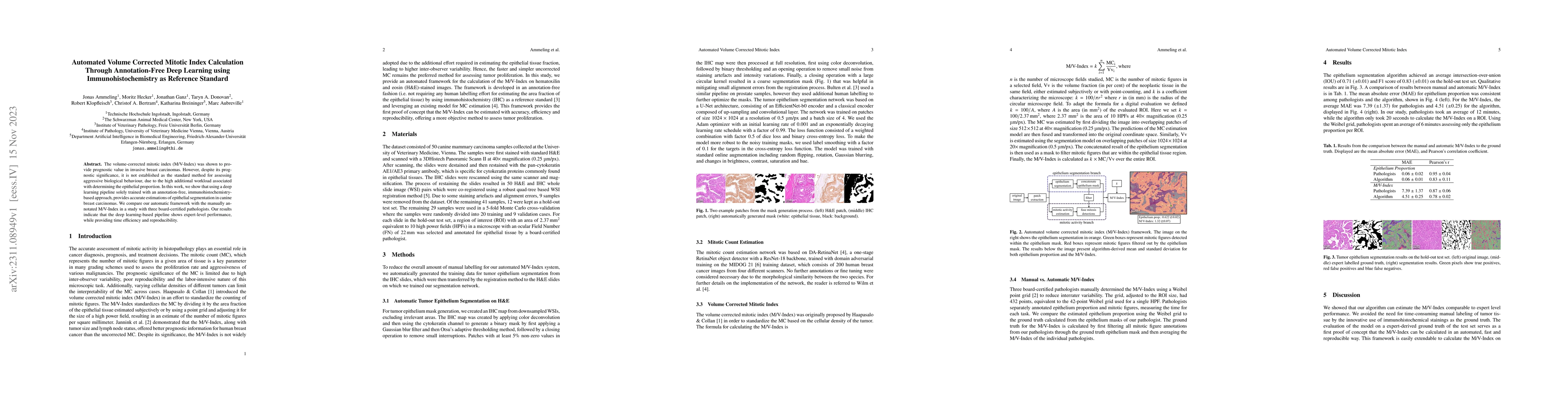
The volume-corrected mitotic index (M/V-Index) was shown to provide prognostic value in invasive breast carcinomas. However, despite its prognostic significance, it is not established as the standar...
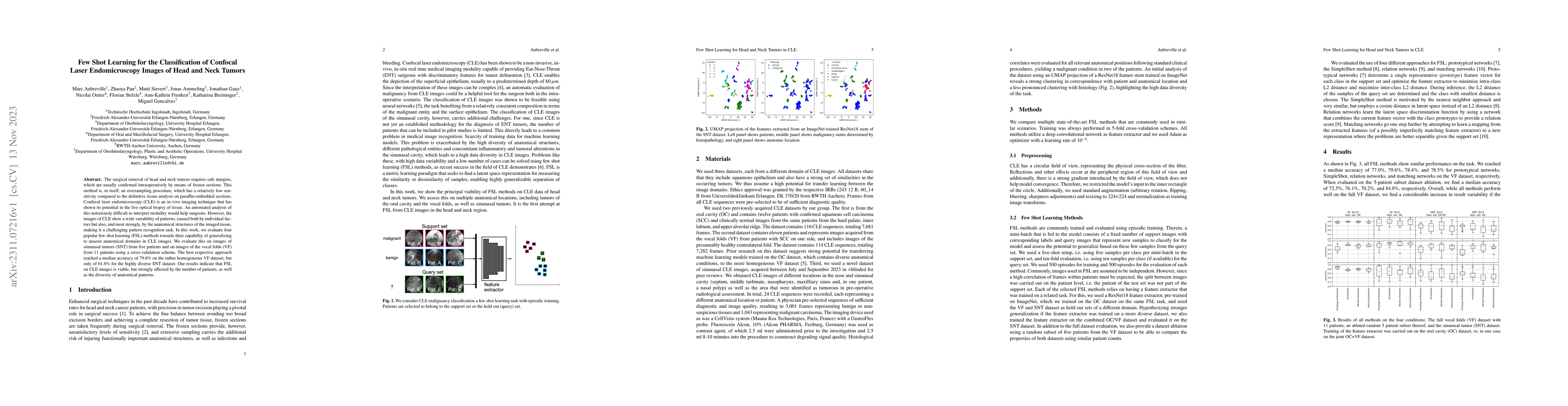
The surgical removal of head and neck tumors requires safe margins, which are usually confirmed intraoperatively by means of frozen sections. This method is, in itself, an oversampling procedure, wh...
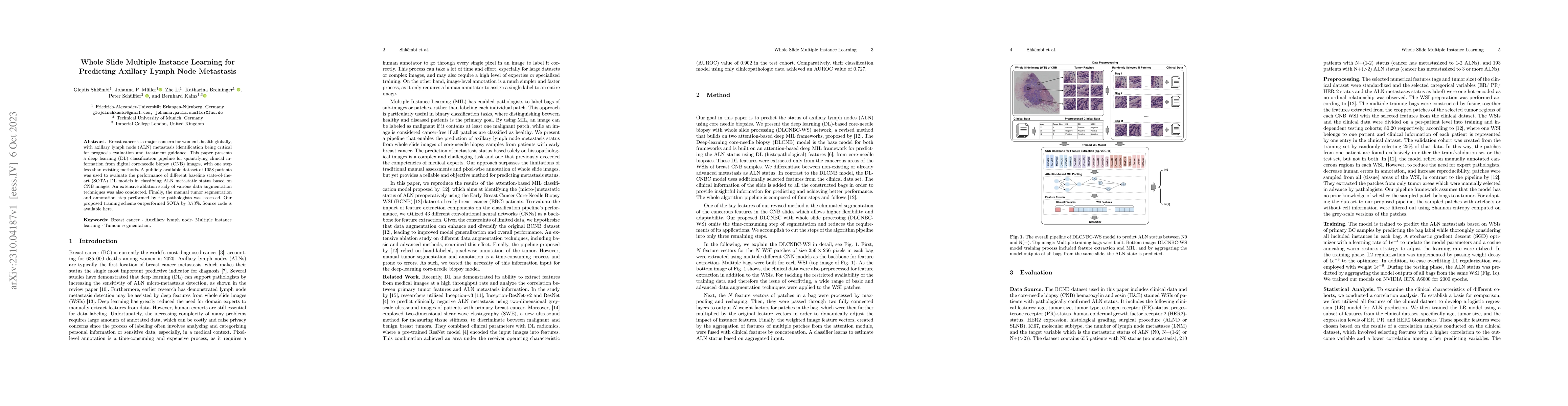
Breast cancer is a major concern for women's health globally, with axillary lymph node (ALN) metastasis identification being critical for prognosis evaluation and treatment guidance. This paper pres...
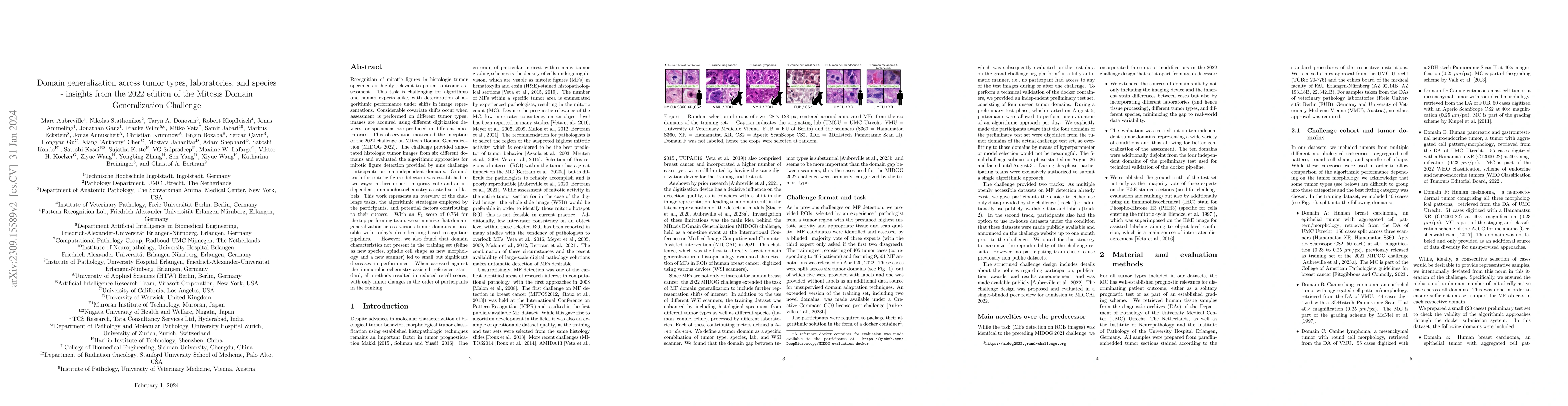
Recognition of mitotic figures in histologic tumor specimens is highly relevant to patient outcome assessment. This task is challenging for algorithms and human experts alike, with deterioration of ...
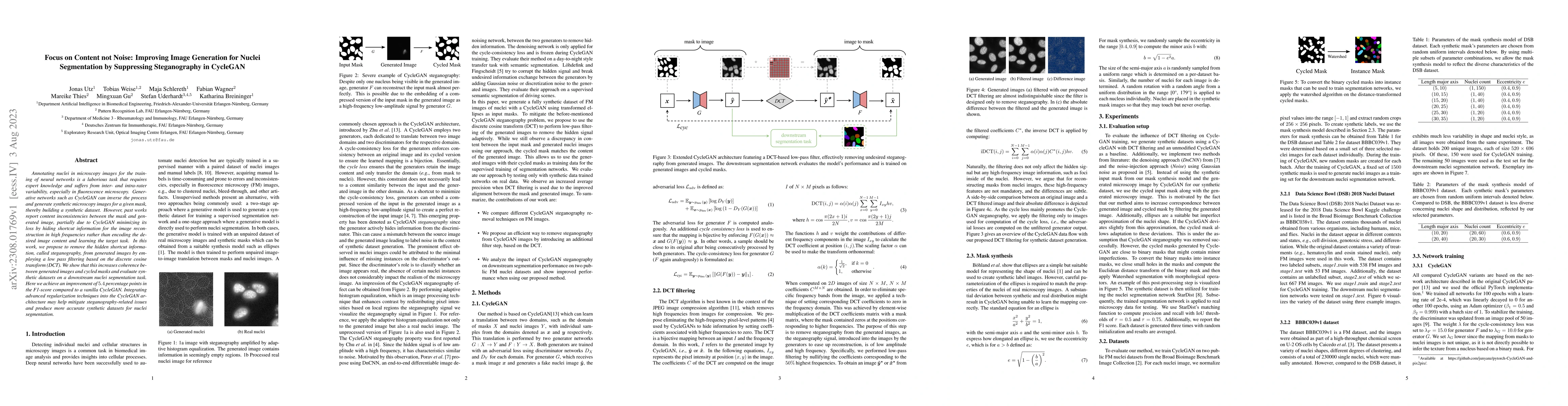
Annotating nuclei in microscopy images for the training of neural networks is a laborious task that requires expert knowledge and suffers from inter- and intra-rater variability, especially in fluor...
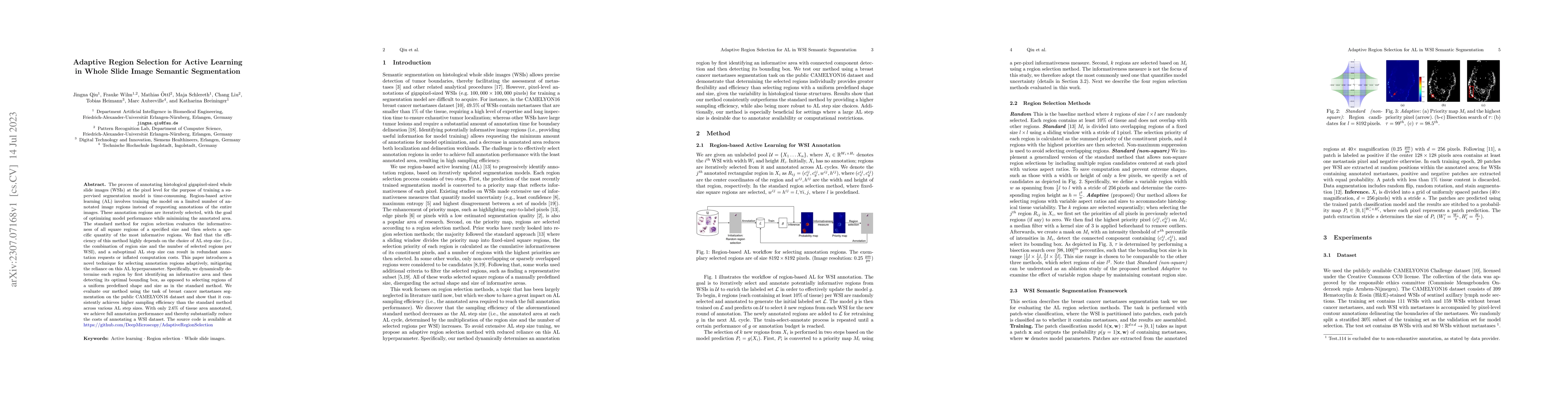
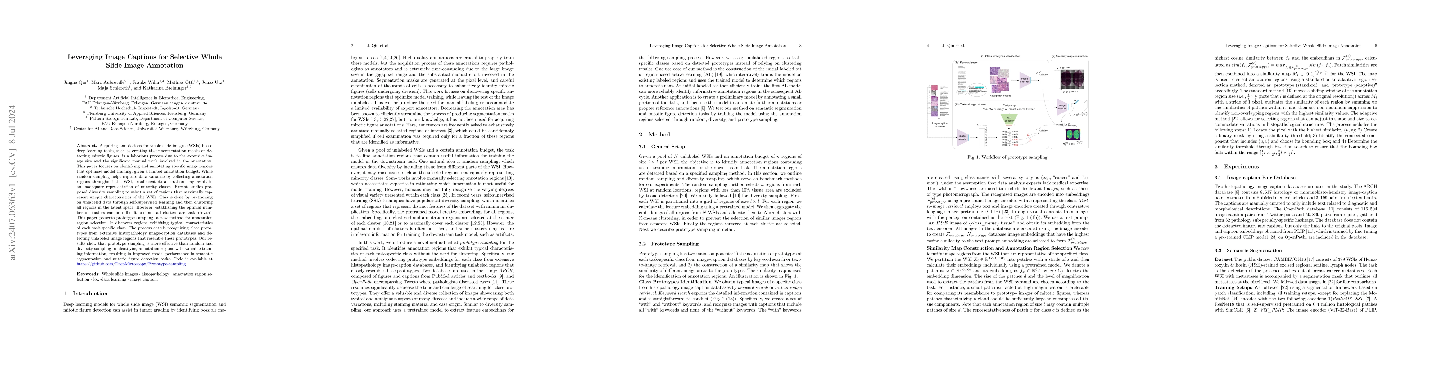
The process of annotating histological gigapixel-sized whole slide images (WSIs) at the pixel level for the purpose of training a supervised segmentation model is time-consuming. Region-based active...
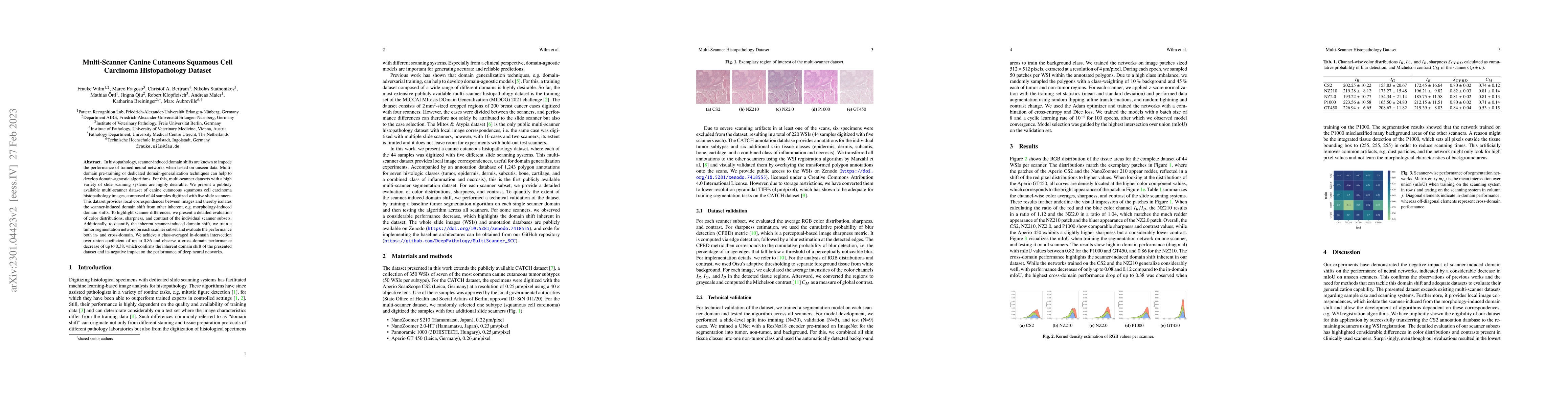
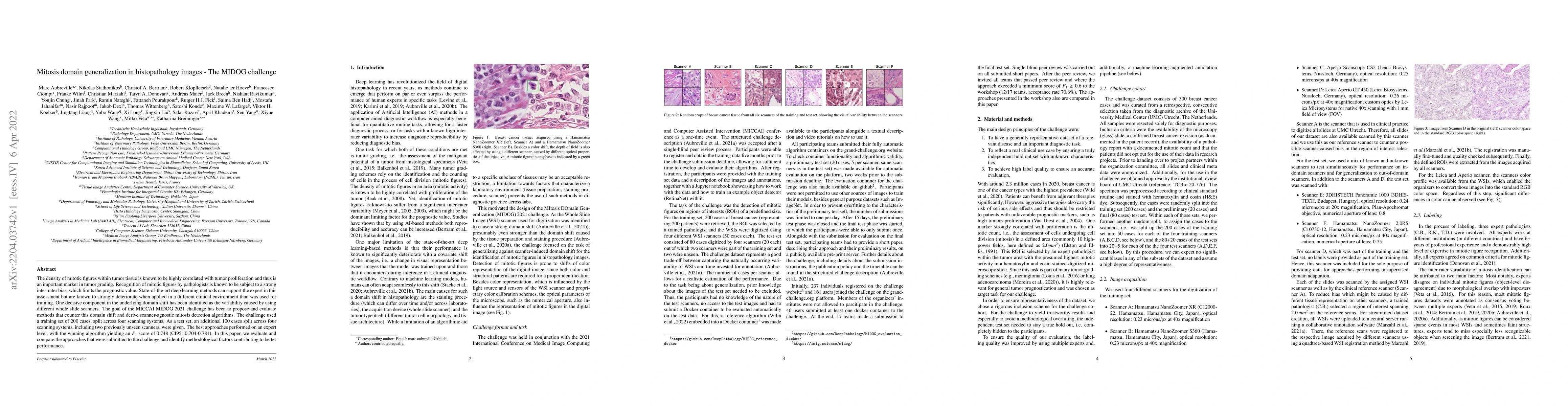
In histopathology, scanner-induced domain shifts are known to impede the performance of trained neural networks when tested on unseen data. Multi-domain pre-training or dedicated domain-generalizati...
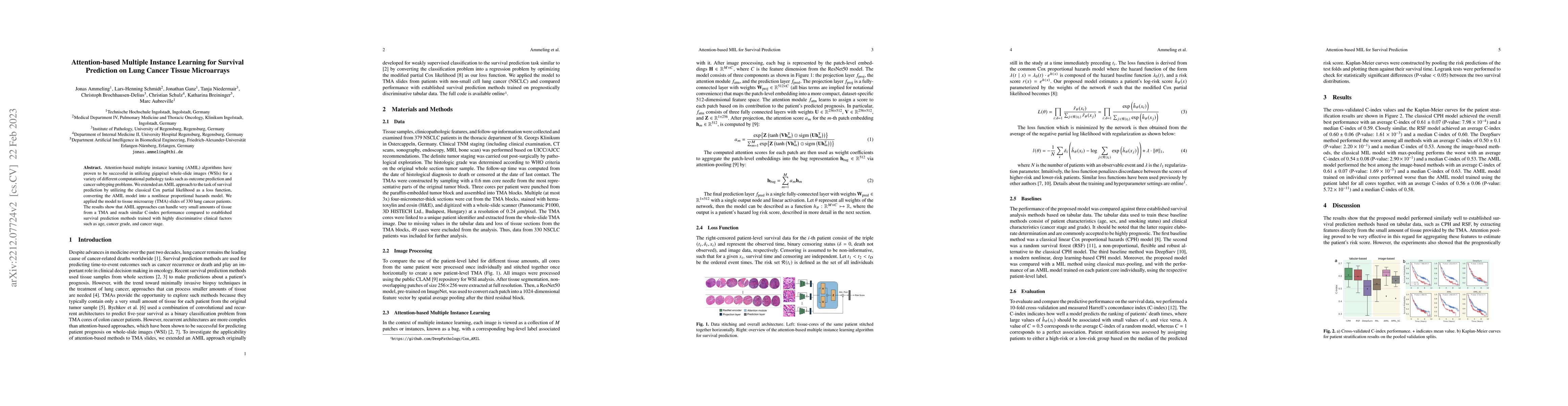
Attention-based multiple instance learning (AMIL) algorithms have proven to be successful in utilizing gigapixel whole-slide images (WSIs) for a variety of different computational pathology tasks su...
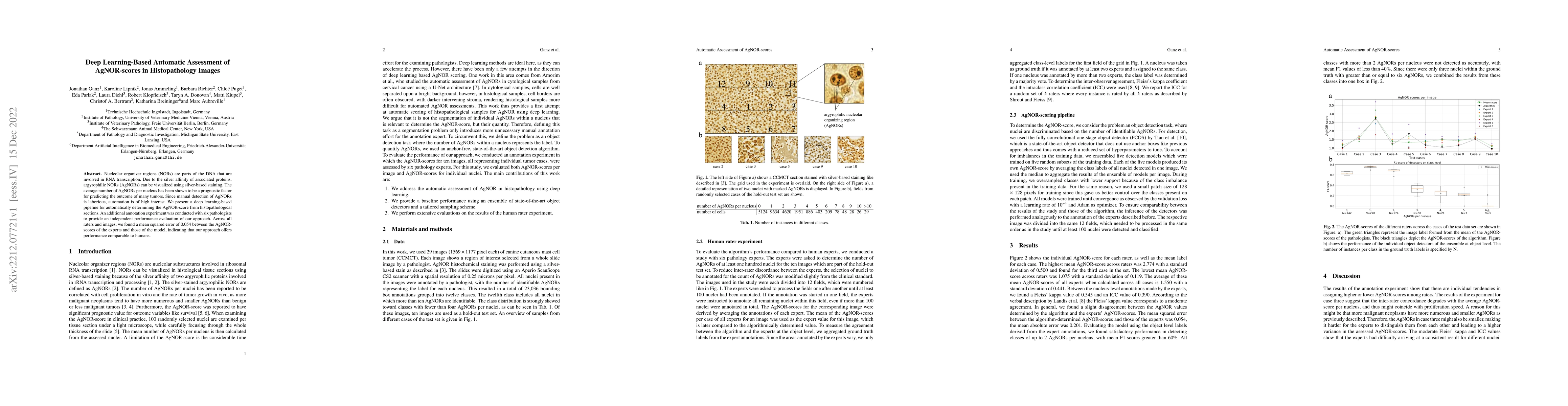
Nucleolar organizer regions (NORs) are parts of the DNA that are involved in RNA transcription. Due to the silver affinity of associated proteins, argyrophilic NORs (AgNORs) can be visualized using ...

Mitotic activity is key for the assessment of malignancy in many tumors. Moreover, it has been demonstrated that the proportion of abnormal mitosis to normal mitosis is of prognostic significance. A...
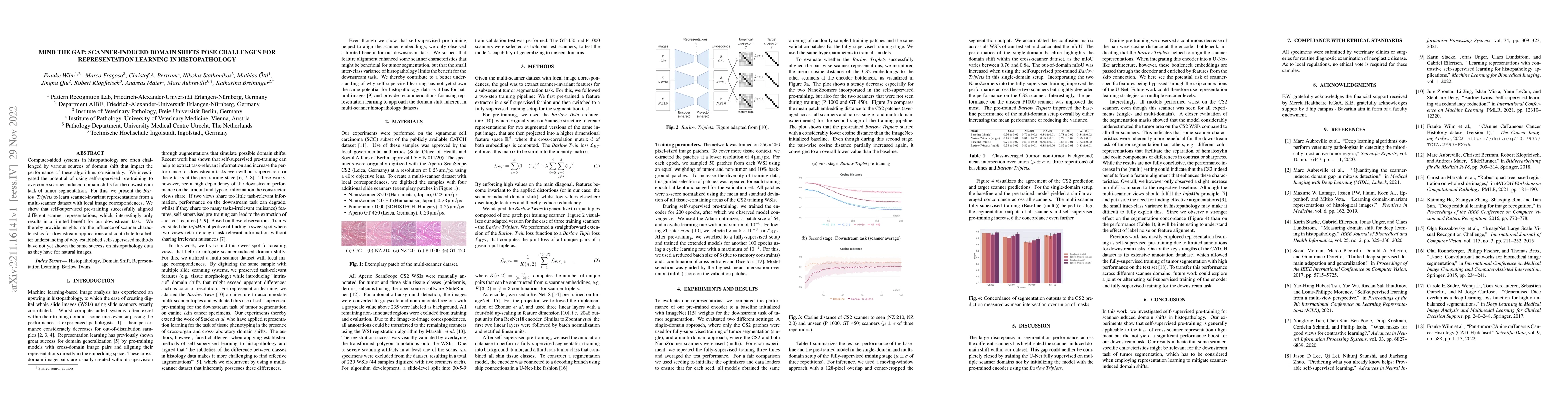
Computer-aided systems in histopathology are often challenged by various sources of domain shift that impact the performance of these algorithms considerably. We investigated the potential of using ...
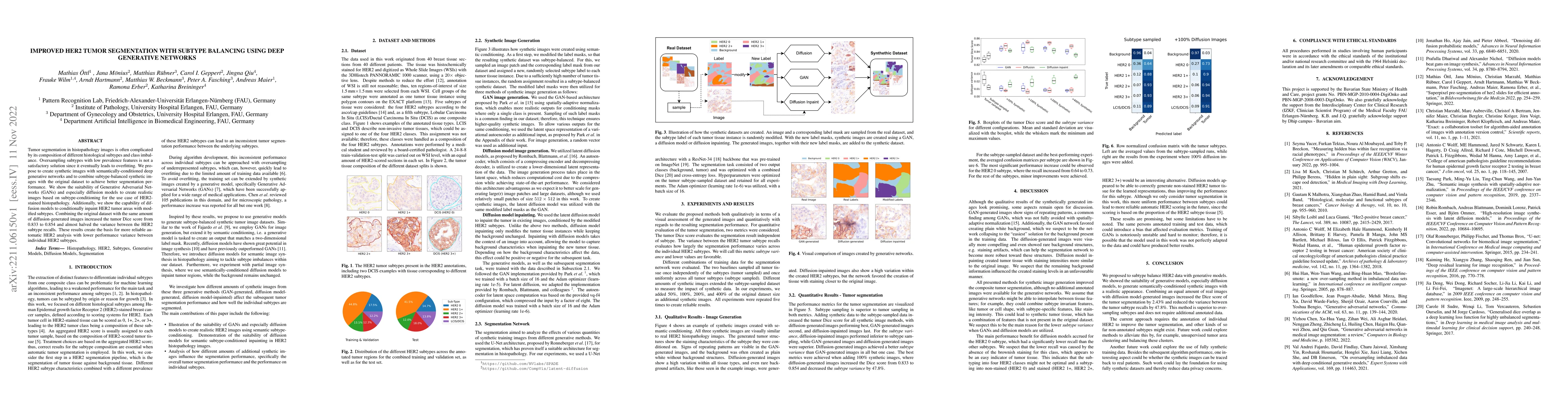
Tumor segmentation in histopathology images is often complicated by its composition of different histological subtypes and class imbalance. Oversampling subtypes with low prevalence features is not ...
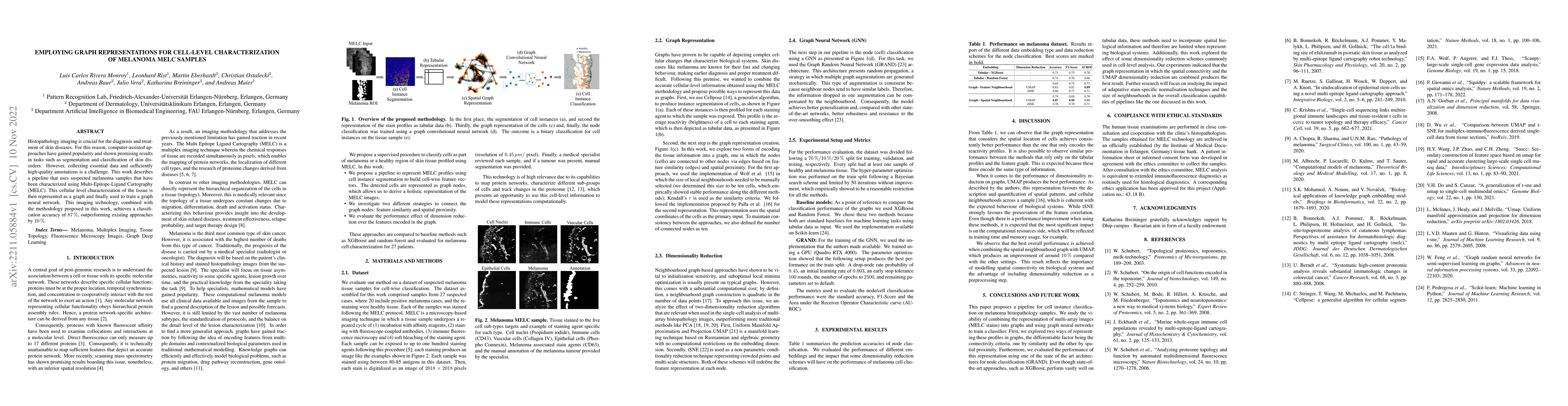
Histopathology imaging is crucial for the diagnosis and treatment of skin diseases. For this reason, computer-assisted approaches have gained popularity and shown promising results in tasks such as ...
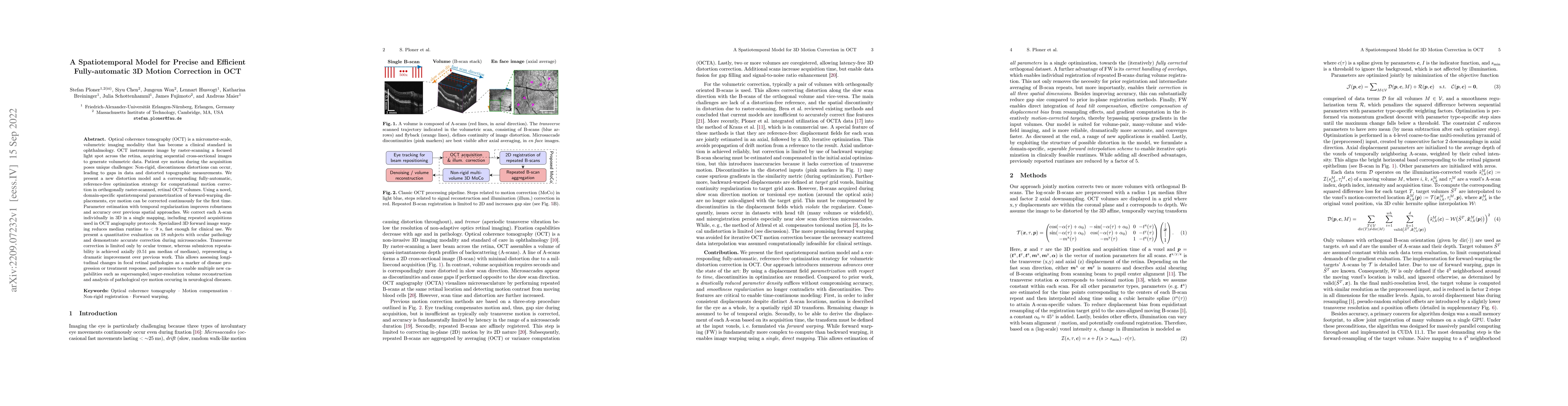
Optical coherence tomography (OCT) is a micrometer-scale, volumetric imaging modality that has become a clinical standard in ophthalmology. OCT instruments image by raster-scanning a focused light s...
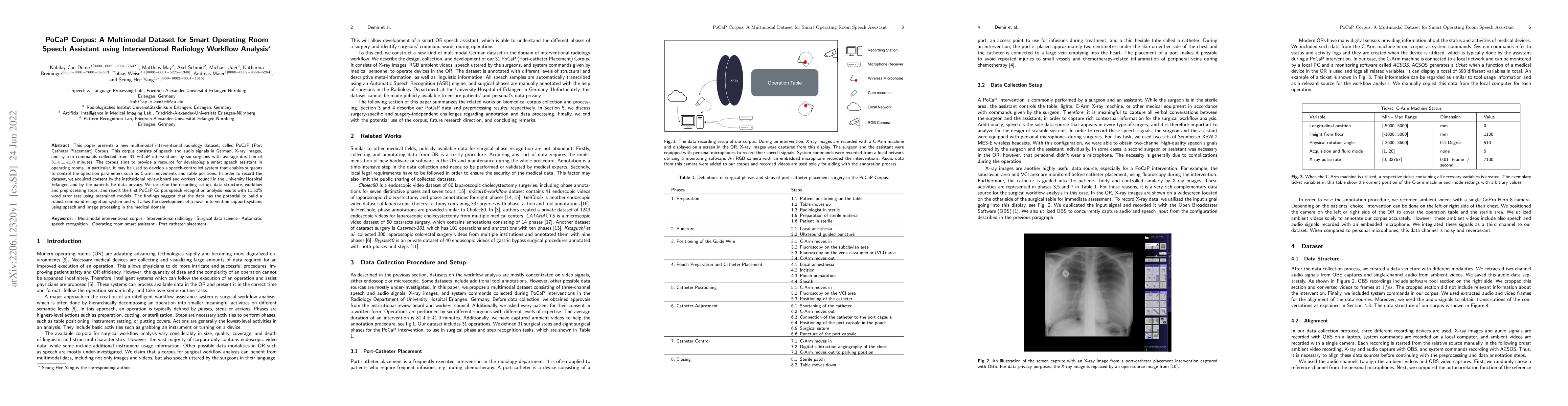
This paper presents a new multimodal interventional radiology dataset, called PoCaP (Port Catheter Placement) Corpus. This corpus consists of speech and audio signals in German, X-ray images, and sy...
The density of mitotic figures within tumor tissue is known to be highly correlated with tumor proliferation and thus is an important marker in tumor grading. Recognition of mitotic figures by patho...

With coronary artery disease (CAD) persisting to be one of the leading causes of death worldwide, interest in supporting physicians with algorithms to speed up and improve diagnosis is high. In clin...
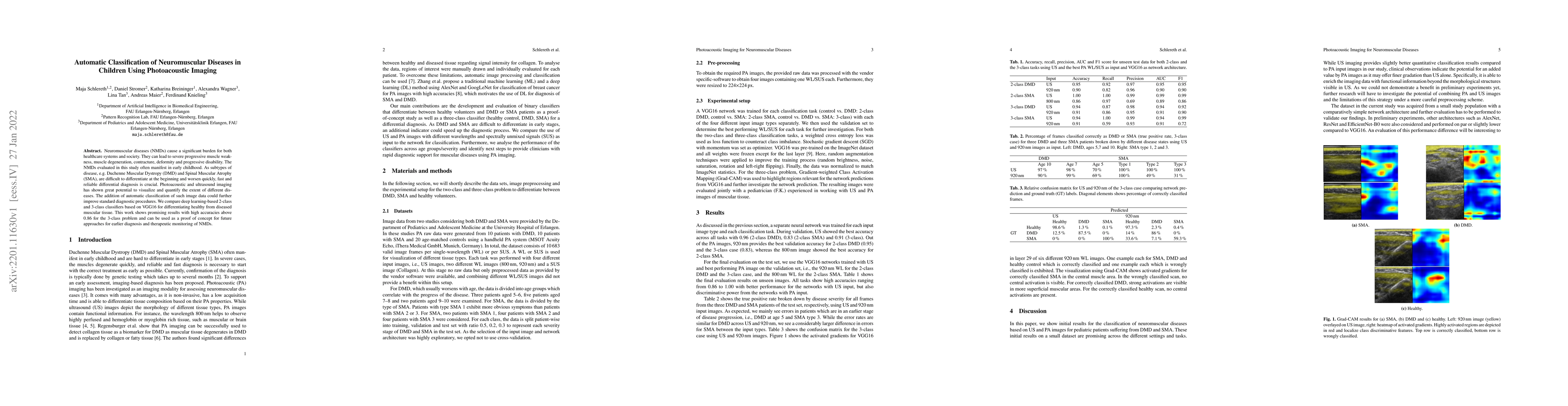
Neuromuscular diseases (NMDs) cause a significant burden for both healthcare systems and society. They can lead to severe progressive muscle weakness, muscle degeneration, contracture, deformity and...
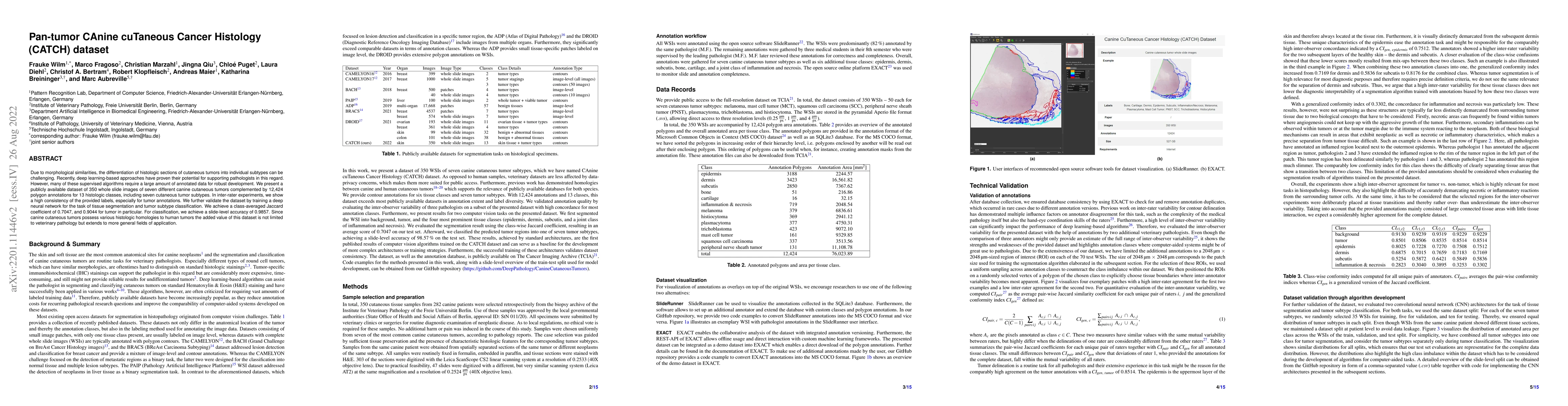
Due to morphological similarities, the differentiation of histologic sections of cutaneous tumors into individual subtypes can be challenging. Recently, deep learning-based approaches have proven th...
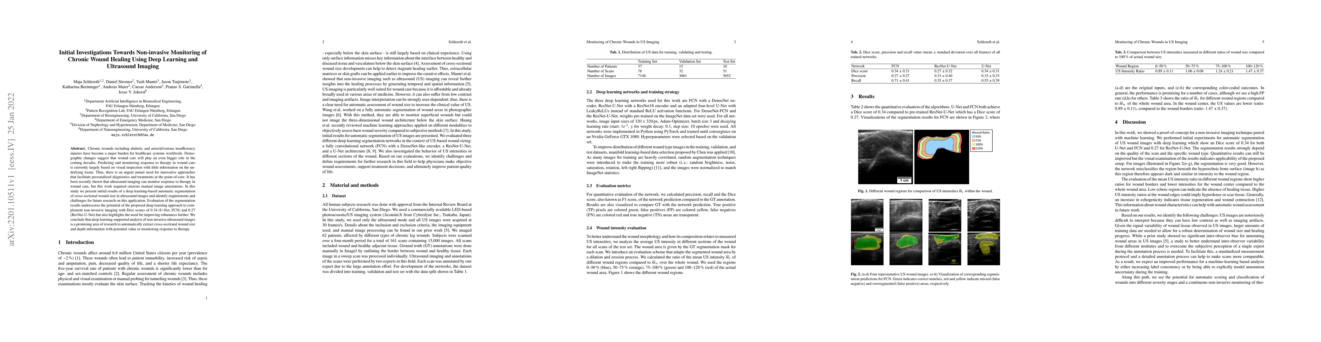
Chronic wounds including diabetic and arterial/venous insufficiency injuries have become a major burden for healthcare systems worldwide. Demographic changes suggest that wound care will play an eve...
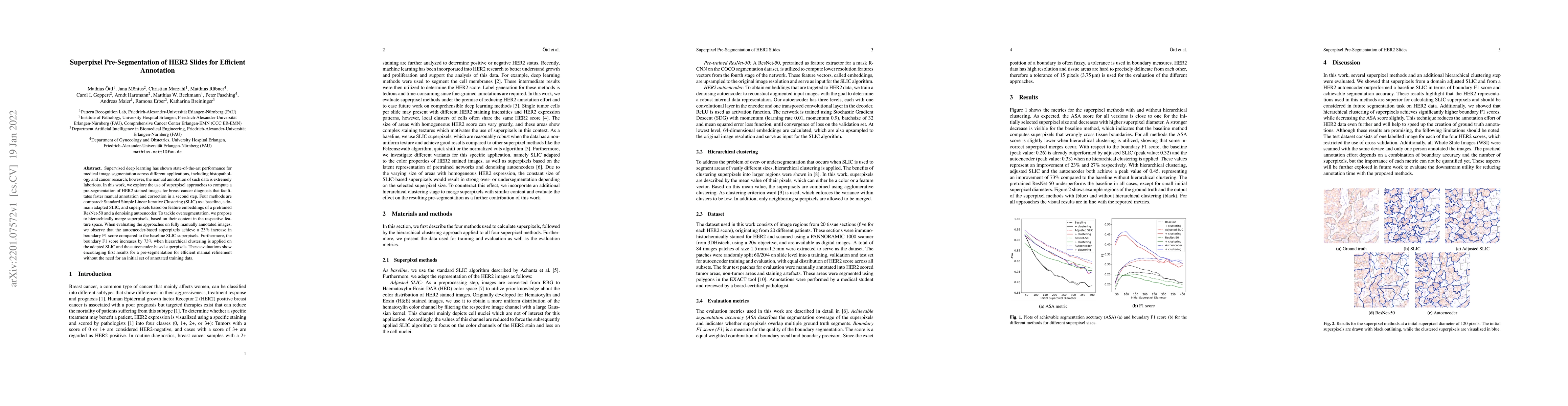
Supervised deep learning has shown state-of-the-art performance for medical image segmentation across different applications, including histopathology and cancer research; however, the manual annota...
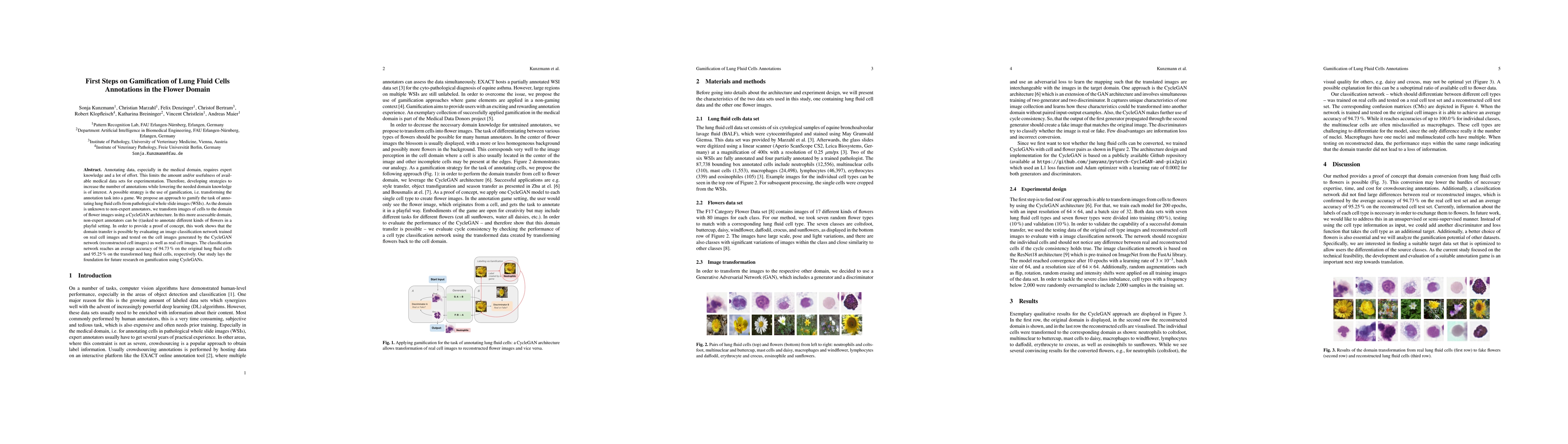
Annotating data, especially in the medical domain, requires expert knowledge and a lot of effort. This limits the amount and/or usefulness of available medical data sets for experimentation. Therefo...
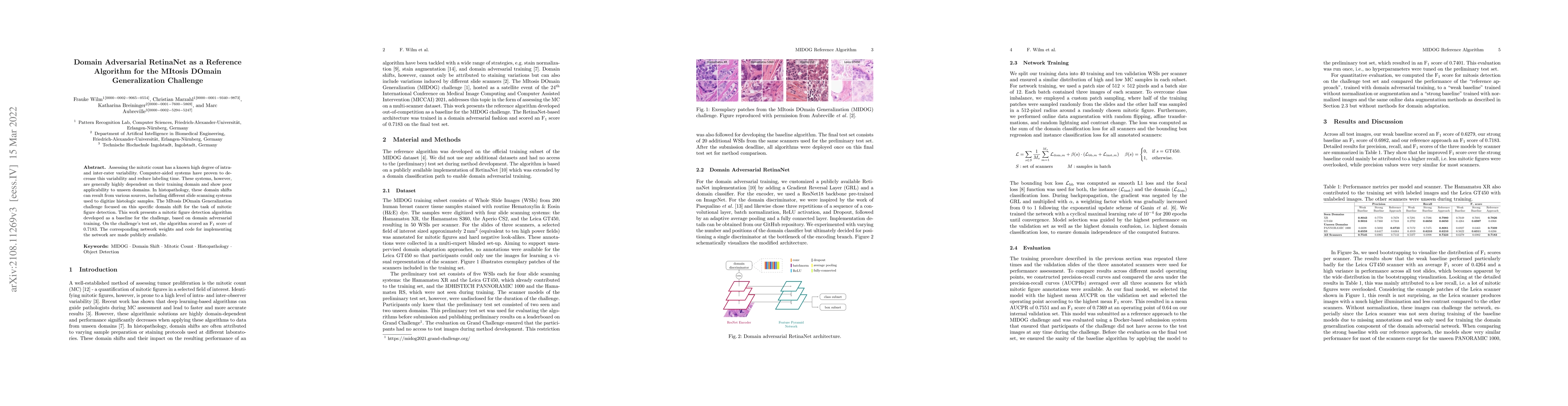
Assessing the Mitotic Count has a known high degree of intra- and inter-rater variability. Computer-aided systems have proven to decrease this variability and reduce labeling time. These systems, ho...
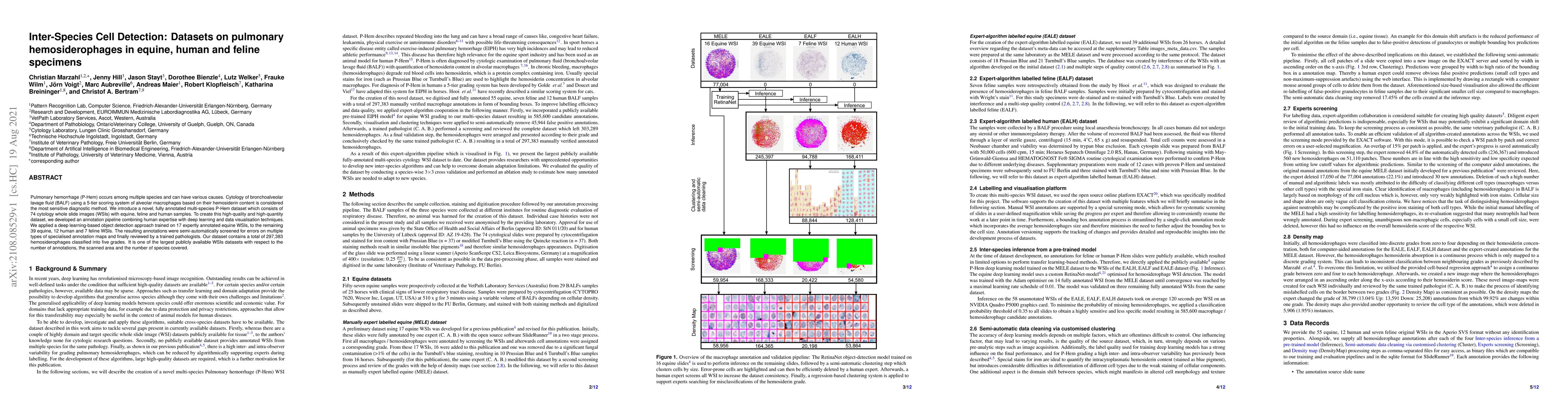
Pulmonary hemorrhage (P-Hem) occurs among multiple species and can have various causes. Cytology of bronchoalveolarlavage fluid (BALF) using a 5-tier scoring system of alveolar macrophages based on ...
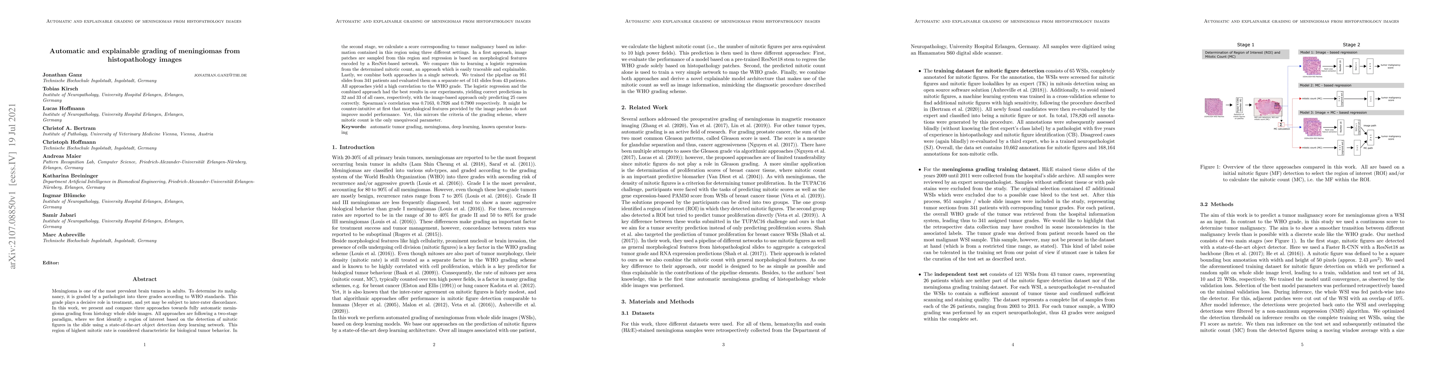
Meningioma is one of the most prevalent brain tumors in adults. To determine its malignancy, it is graded by a pathologist into three grades according to WHO standards. This grade plays a decisive r...
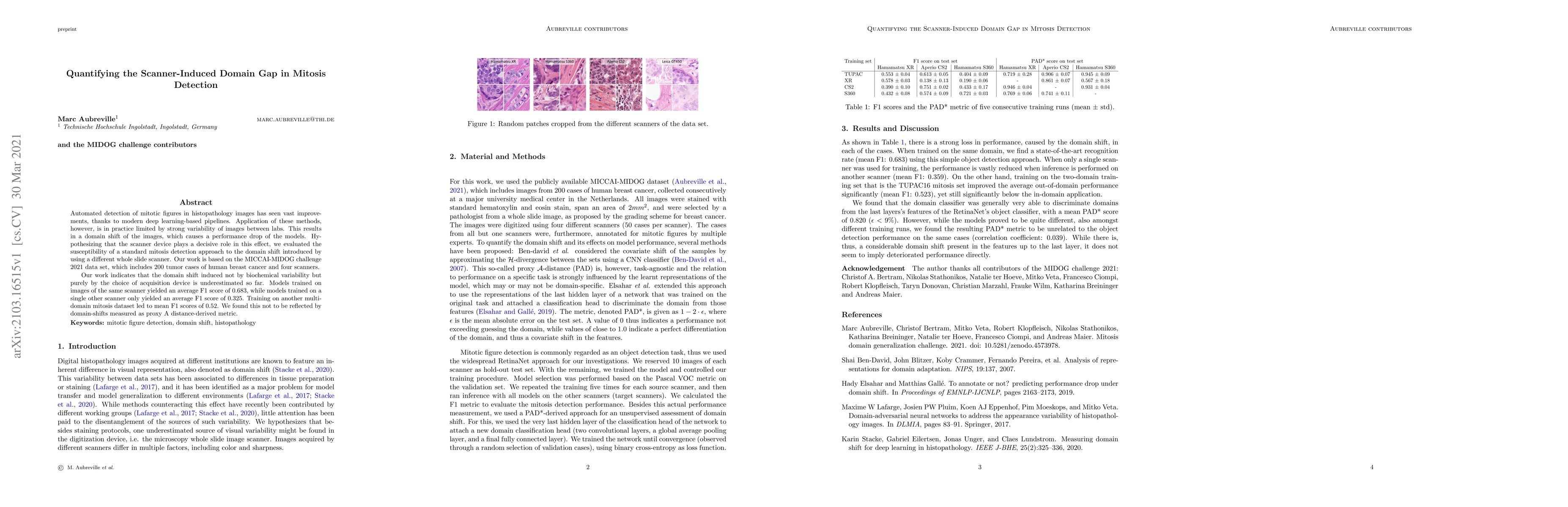
Automated detection of mitotic figures in histopathology images has seen vast improvements, thanks to modern deep learning-based pipelines. Application of these methods, however, is in practice limi...
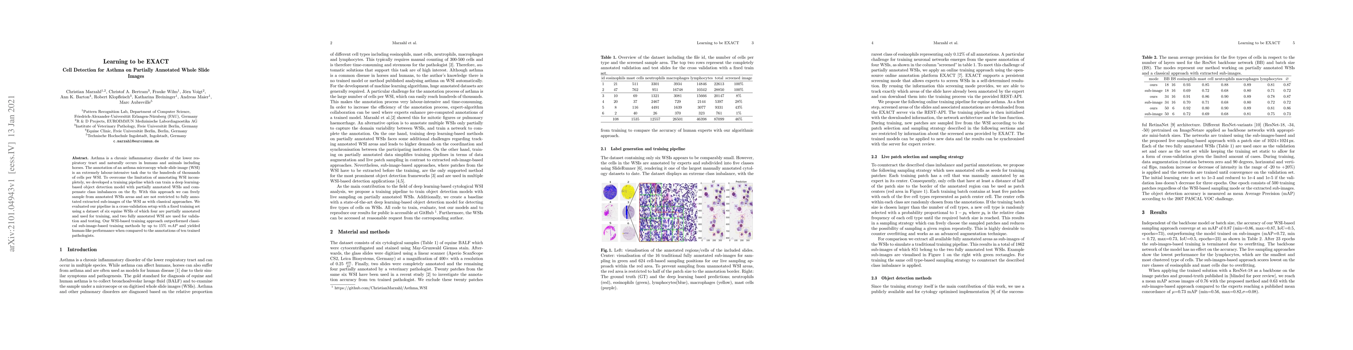
Asthma is a chronic inflammatory disorder of the lower respiratory tract and naturally occurs in humans and animals including horses. The annotation of an asthma microscopy whole slide image (WSI) i...
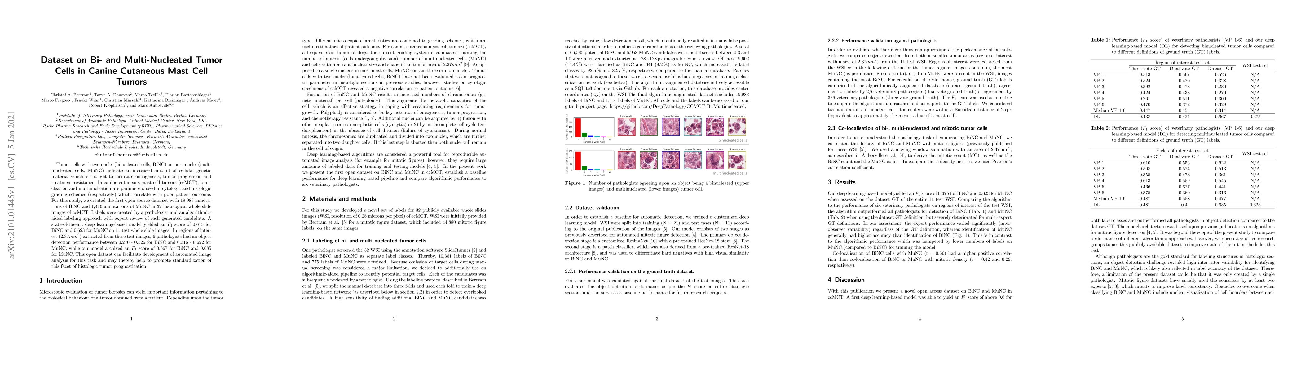
Tumor cells with two nuclei (binucleated cells, BiNC) or more nuclei (multinucleated cells, MuNC) indicate an increased amount of cellular genetic material which is thought to facilitate oncogenesis...
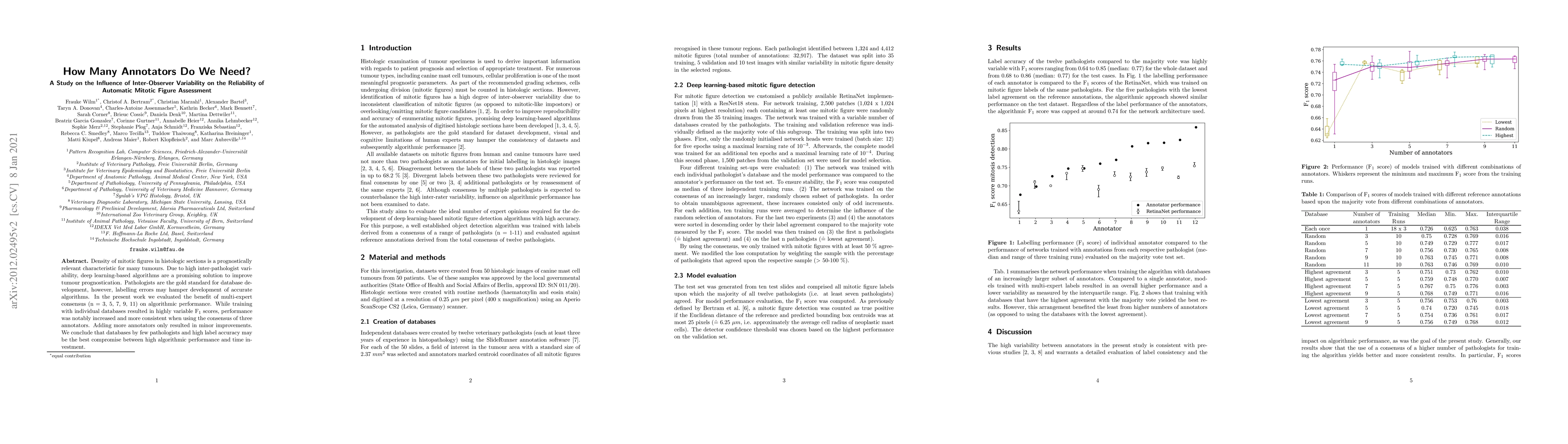
Density of mitotic figures in histologic sections is a prognostically relevant characteristic for many tumours. Due to high inter-pathologist variability, deep learning-based algorithms are a promis...
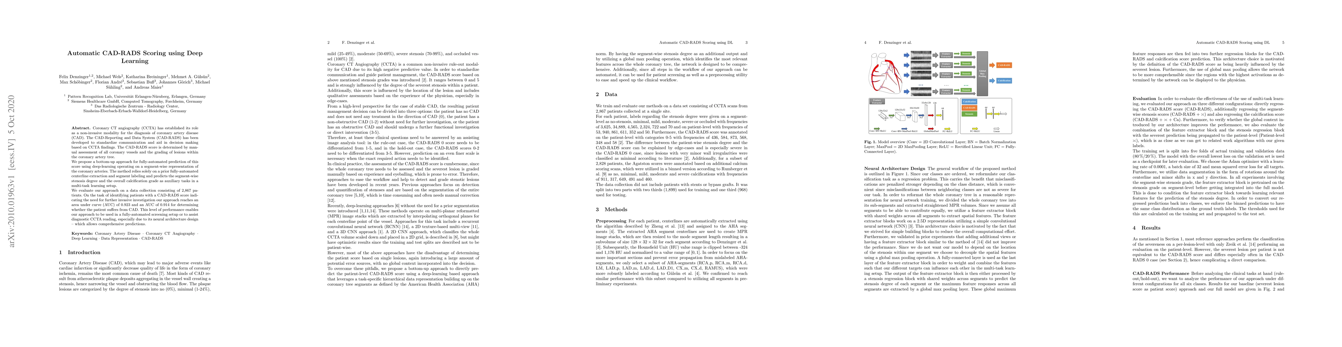
Coronary CT angiography (CCTA) has established its role as a non-invasive modality for the diagnosis of coronary artery disease (CAD). The CAD-Reporting and Data System (CAD-RADS) has been developed...
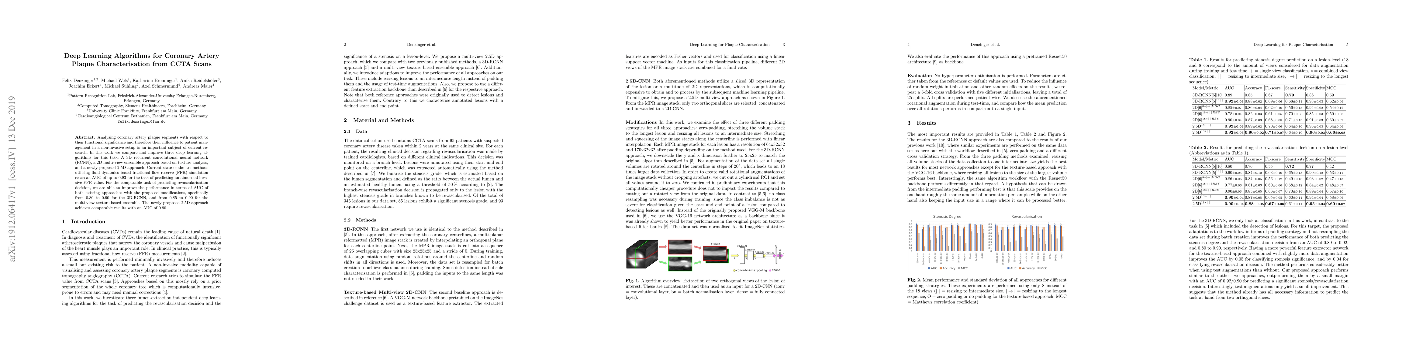
Analysing coronary artery plaque segments with respect to their functional significance and therefore their influence to patient management in a non-invasive setup is an important subject of current...
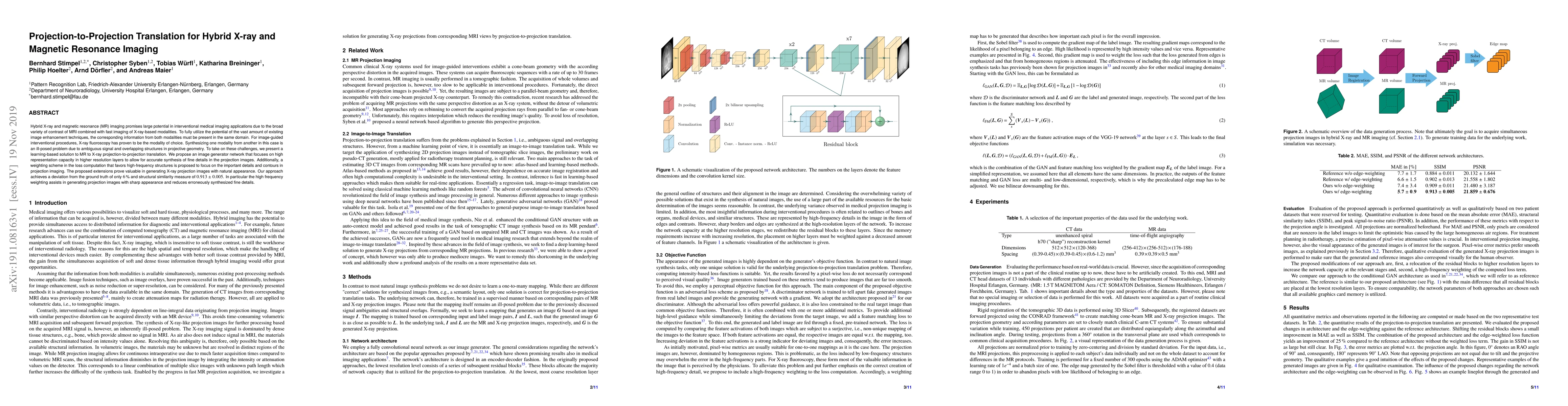
Hybrid X-ray and magnetic resonance (MR) imaging promises large potential in interventional medical imaging applications due to the broad variety of contrast of MRI combined with fast imaging of X-r...
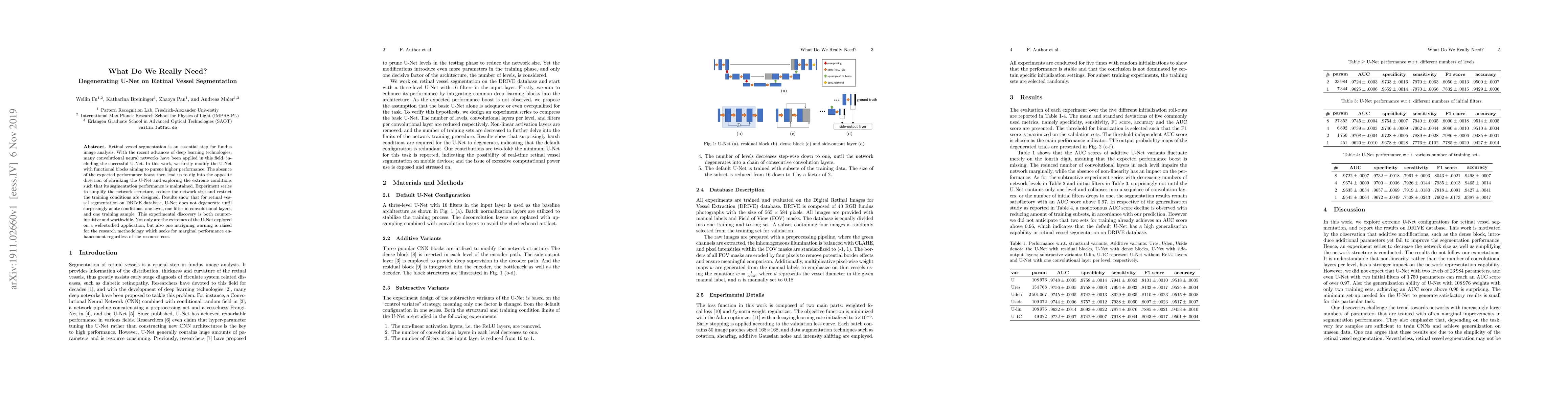
Retinal vessel segmentation is an essential step for fundus image analysis. With the recent advances of deep learning technologies, many convolutional neural networks have been applied in this field...
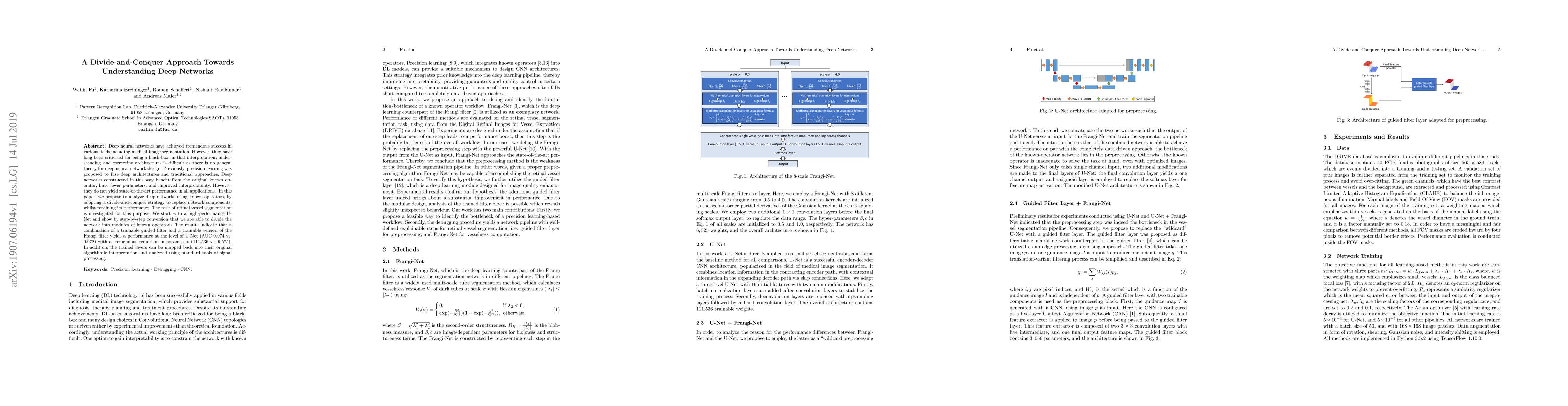
Deep neural networks have achieved tremendous success in various fields including medical image segmentation. However, they have long been criticized for being a black-box, in that interpretation, u...
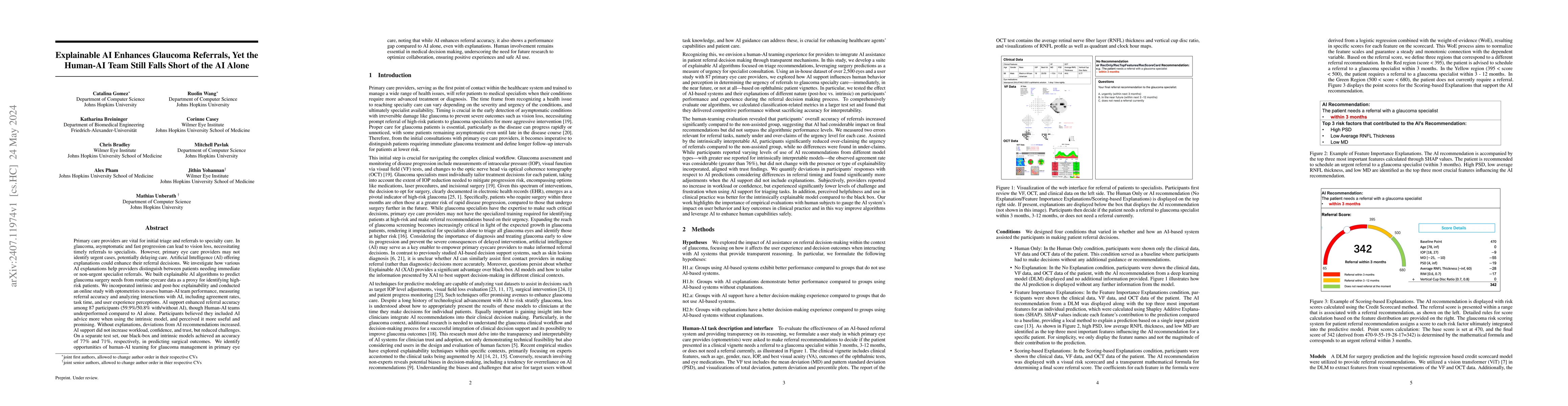
Primary care providers are vital for initial triage and referrals to specialty care. In glaucoma, asymptomatic and fast progression can lead to vision loss, necessitating timely referrals to specialis...
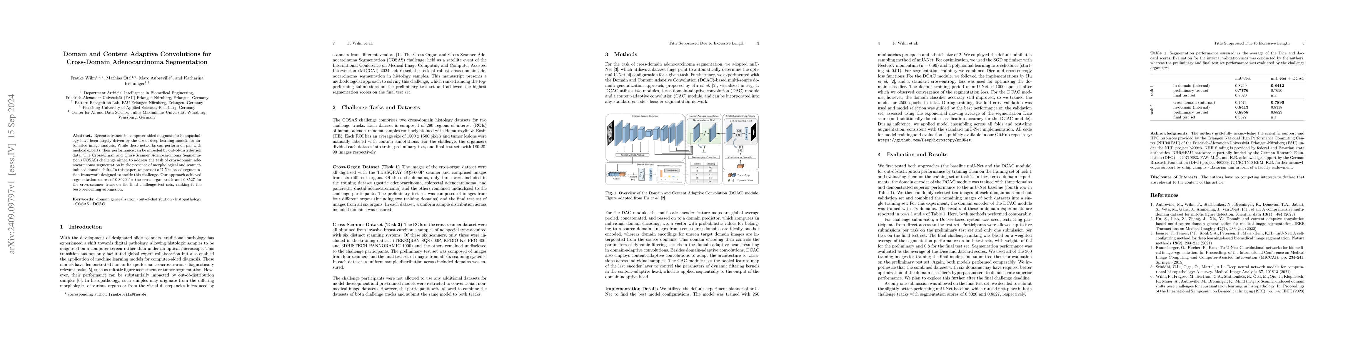
Recent advances in computer-aided diagnosis for histopathology have been largely driven by the use of deep learning models for automated image analysis. While these networks can perform on par with me...
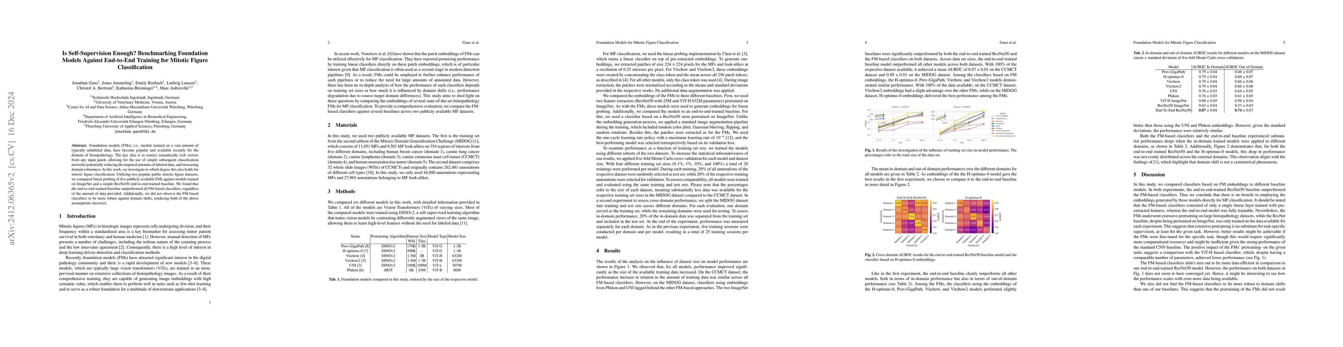
Foundation models (FMs), i.e., models trained on a vast amount of typically unlabeled data, have become popular and available recently for the domain of histopathology. The key idea is to extract sema...
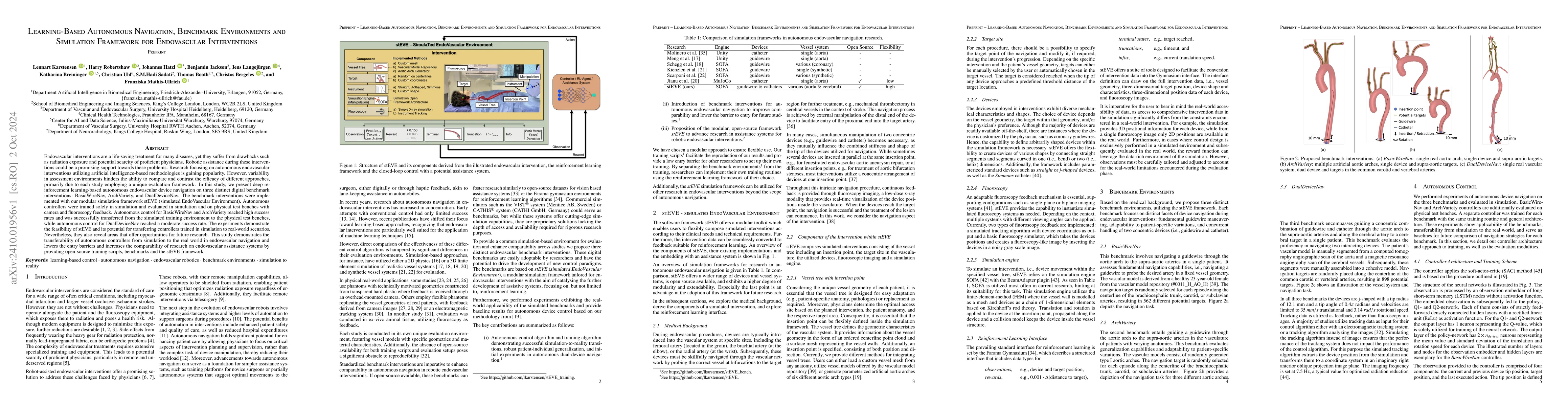
Endovascular interventions are a life-saving treatment for many diseases, yet suffer from drawbacks such as radiation exposure and potential scarcity of proficient physicians. Robotic assistance durin...
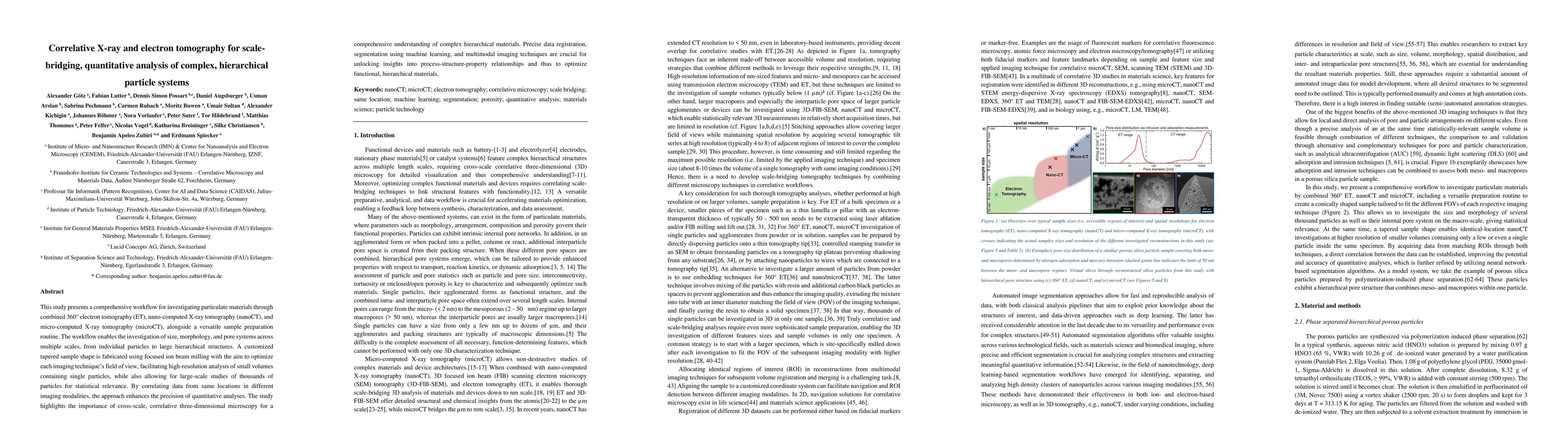
This study presents a comprehensive workflow for investigating particulate materials through combined 360{\deg} electron tomography (ET), nano-computed X-ray tomography (nanoCT), and micro-computed X-...
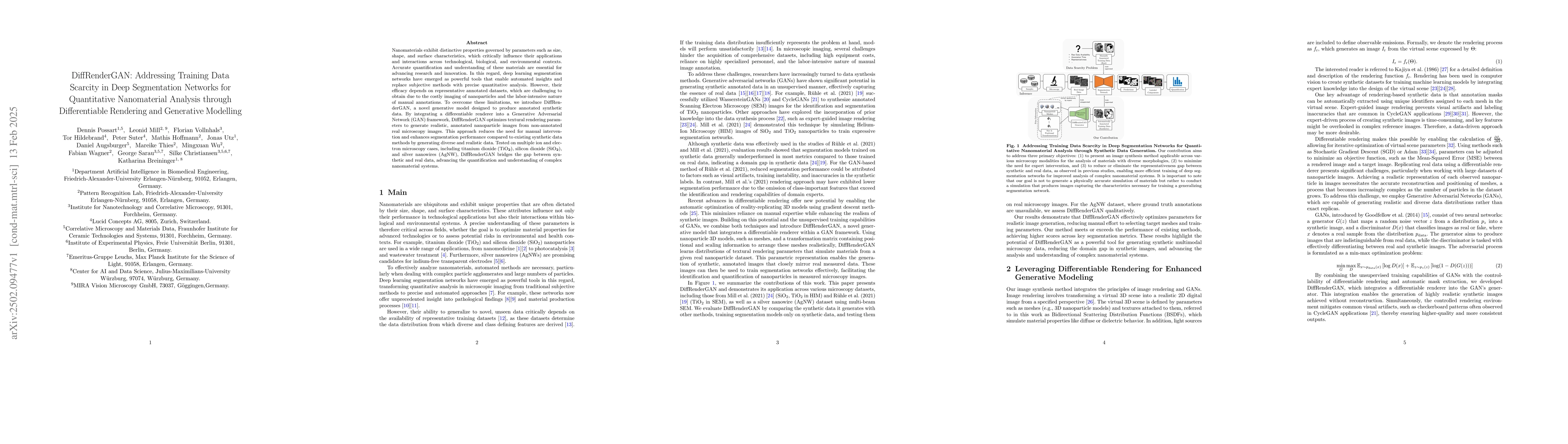
Nanomaterials exhibit distinctive properties governed by parameters such as size, shape, and surface characteristics, which critically influence their applications and interactions across technologica...
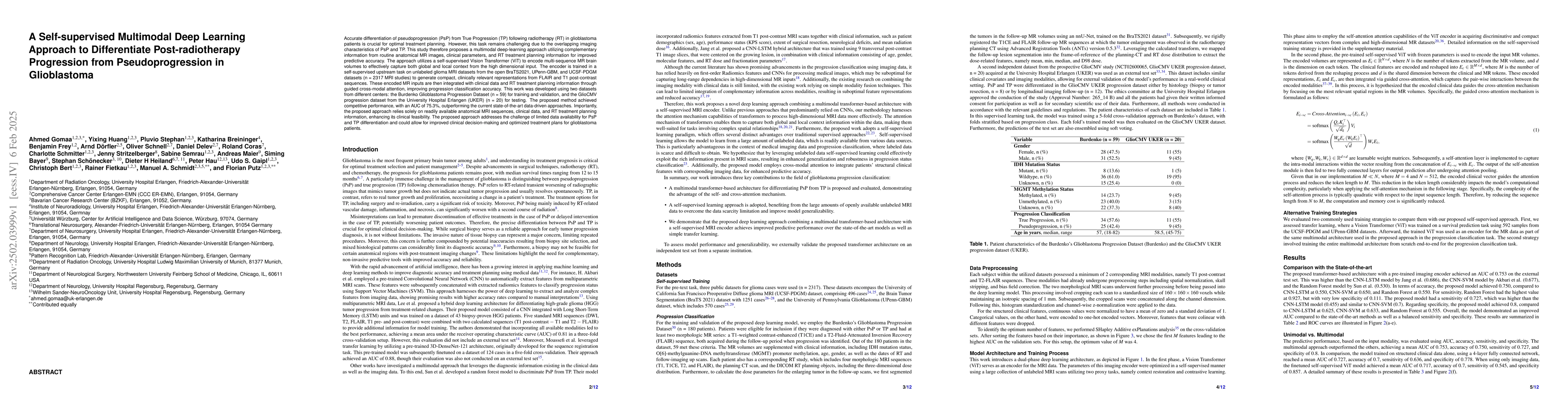
Accurate differentiation of pseudoprogression (PsP) from True Progression (TP) following radiotherapy (RT) in glioblastoma (GBM) patients is crucial for optimal treatment planning. However, this task ...
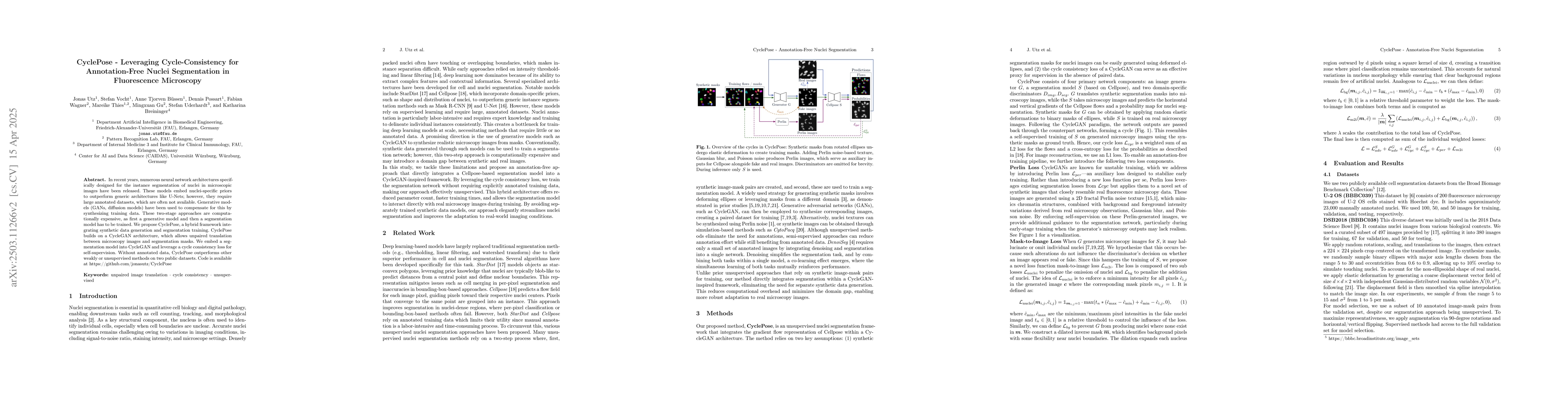
In recent years, numerous neural network architectures specifically designed for the instance segmentation of nuclei in microscopic images have been released. These models embed nuclei-specific priors...
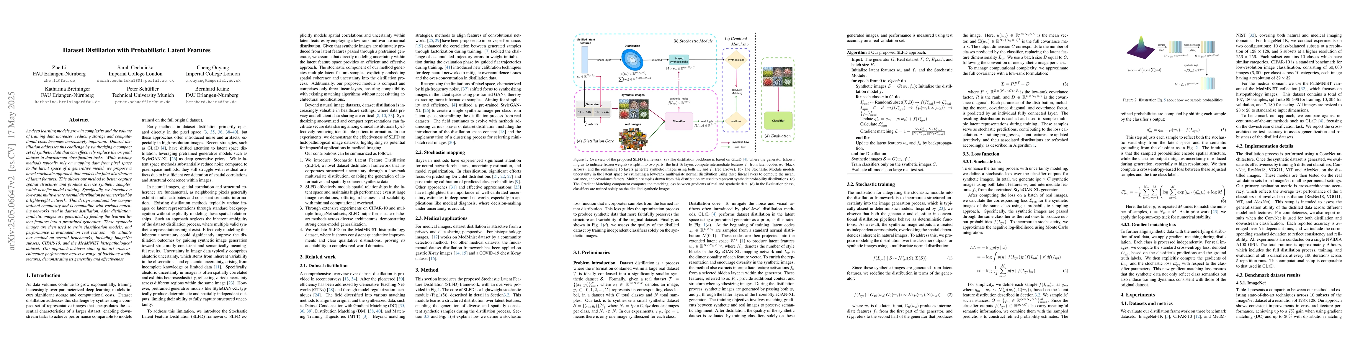
As deep learning models grow in complexity and the volume of training data increases, reducing storage and computational costs becomes increasingly important. Dataset distillation addresses this chall...
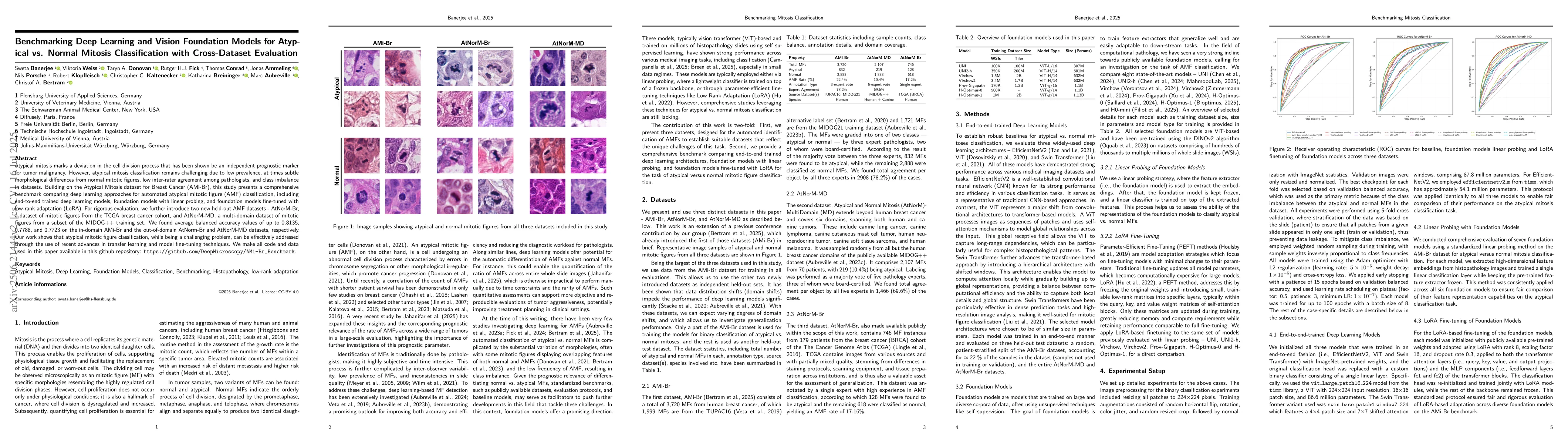
Atypical mitoses mark a deviation in the cell division process that can be an independent prognostically relevant marker for tumor malignancy. However, their identification remains challenging due to ...
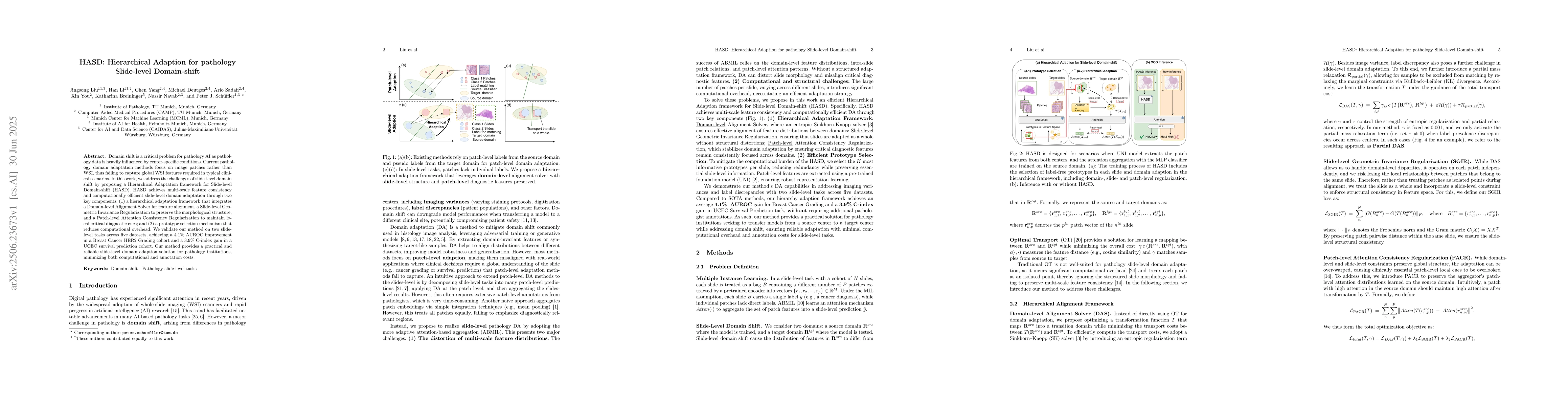
Domain shift is a critical problem for pathology AI as pathology data is heavily influenced by center-specific conditions. Current pathology domain adaptation methods focus on image patches rather tha...
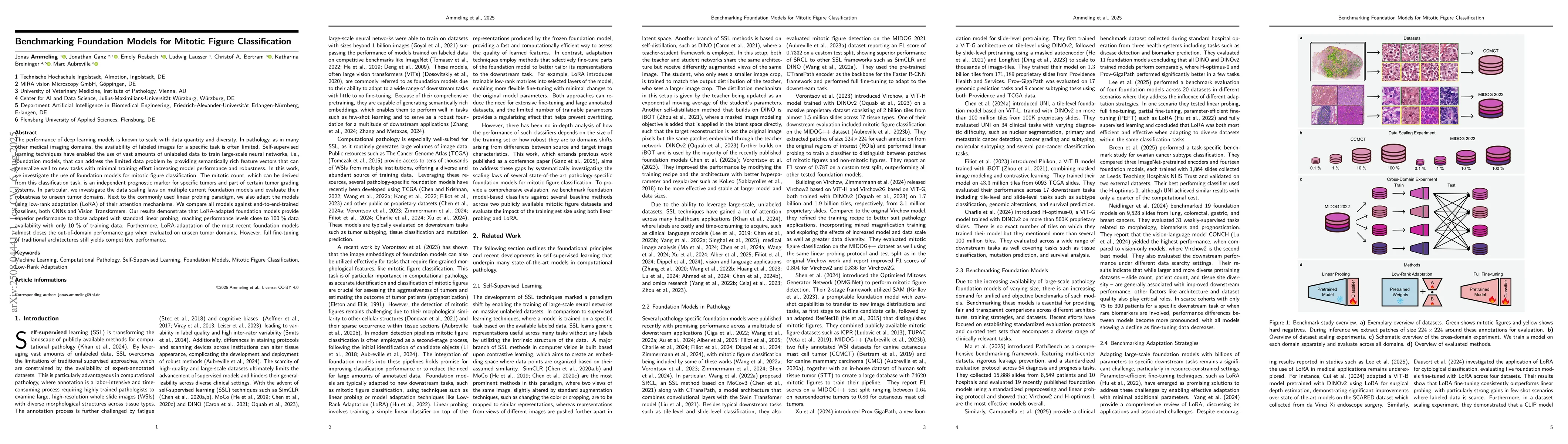
The performance of deep learning models is known to scale with data quantity and diversity. In pathology, as in many other medical imaging domains, the availability of labeled images for a specific ta...
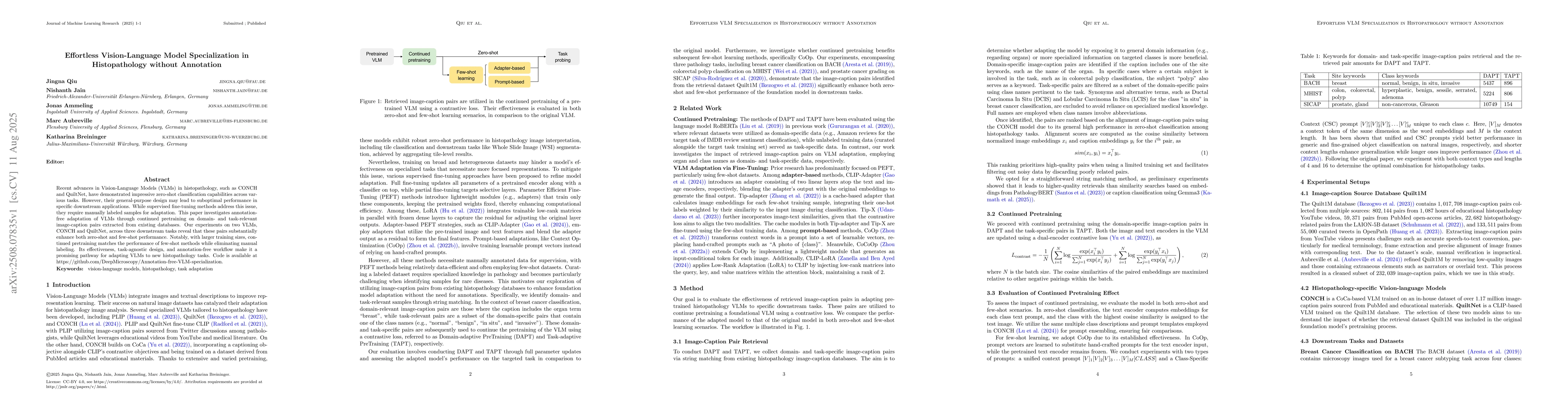
Recent advances in Vision-Language Models (VLMs) in histopathology, such as CONCH and QuiltNet, have demonstrated impressive zero-shot classification capabilities across various tasks. However, their ...
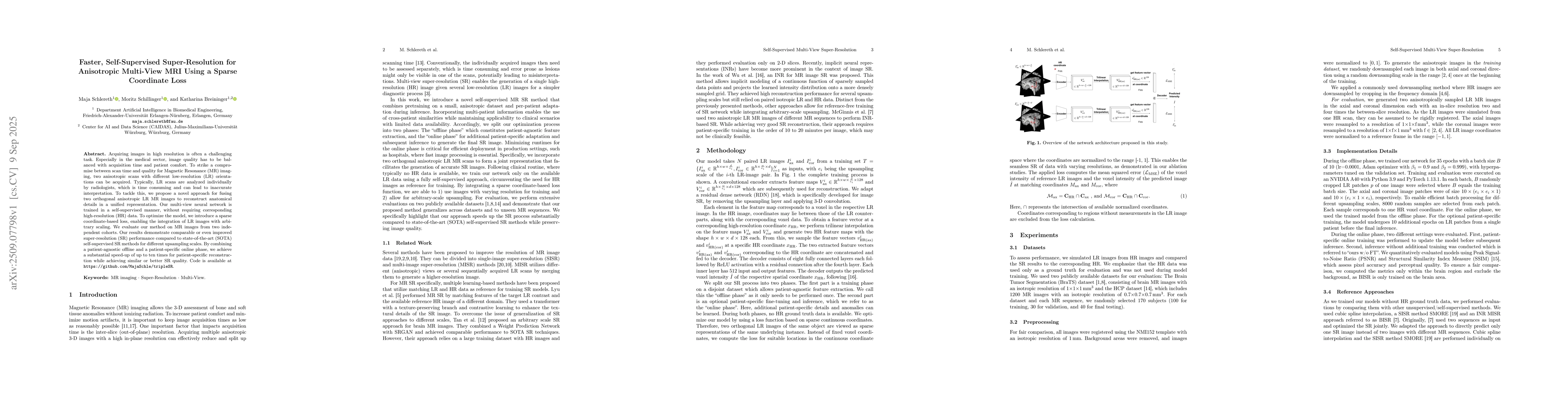
Acquiring images in high resolution is often a challenging task. Especially in the medical sector, image quality has to be balanced with acquisition time and patient comfort. To strike a compromise be...
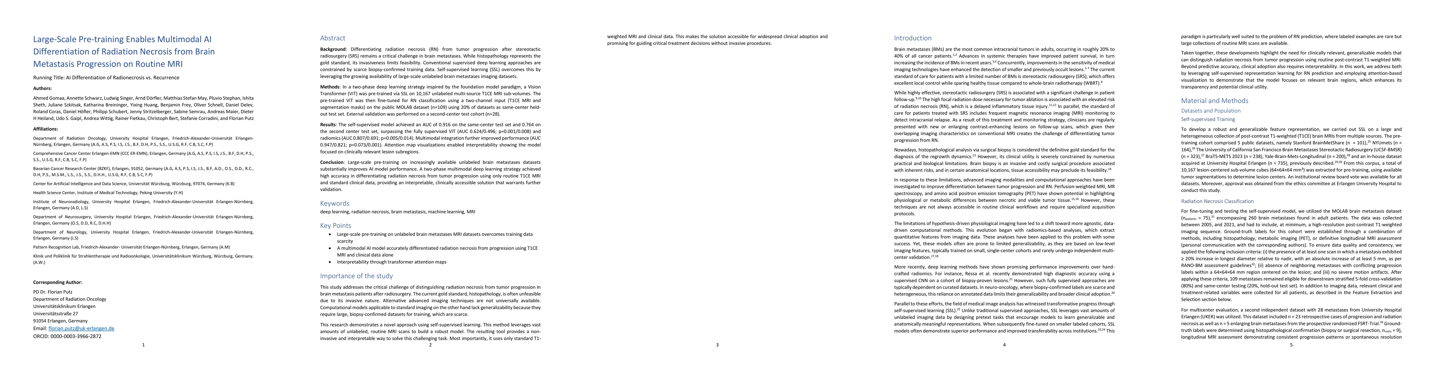
Background: Differentiating radiation necrosis (RN) from tumor progression after stereotactic radiosurgery (SRS) remains a critical challenge in brain metastases. While histopathology represents the g...
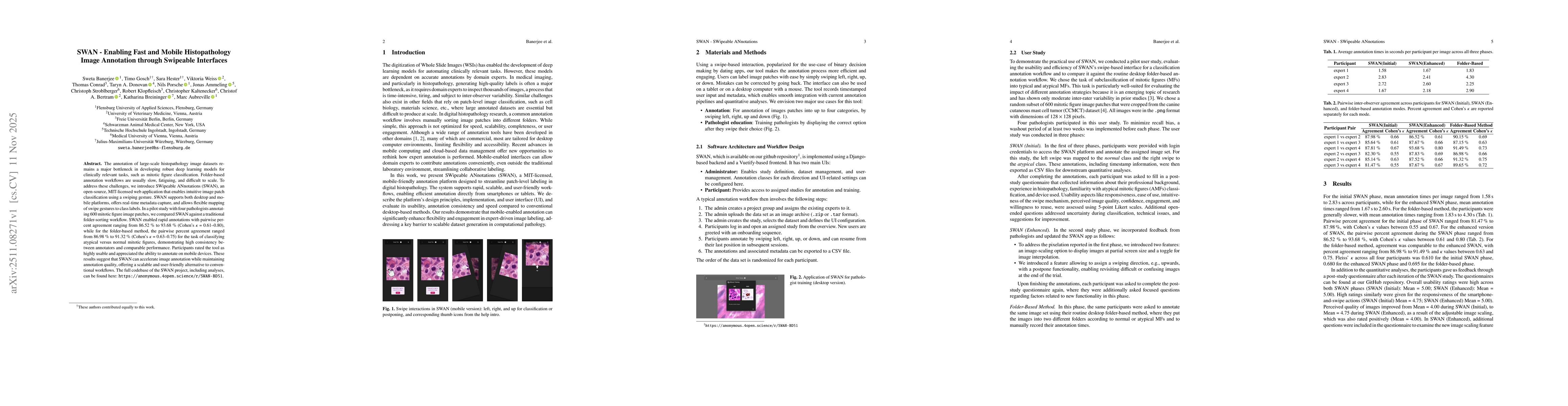
The annotation of large scale histopathology image datasets remains a major bottleneck in developing robust deep learning models for clinically relevant tasks, such as mitotic figure classification. F...
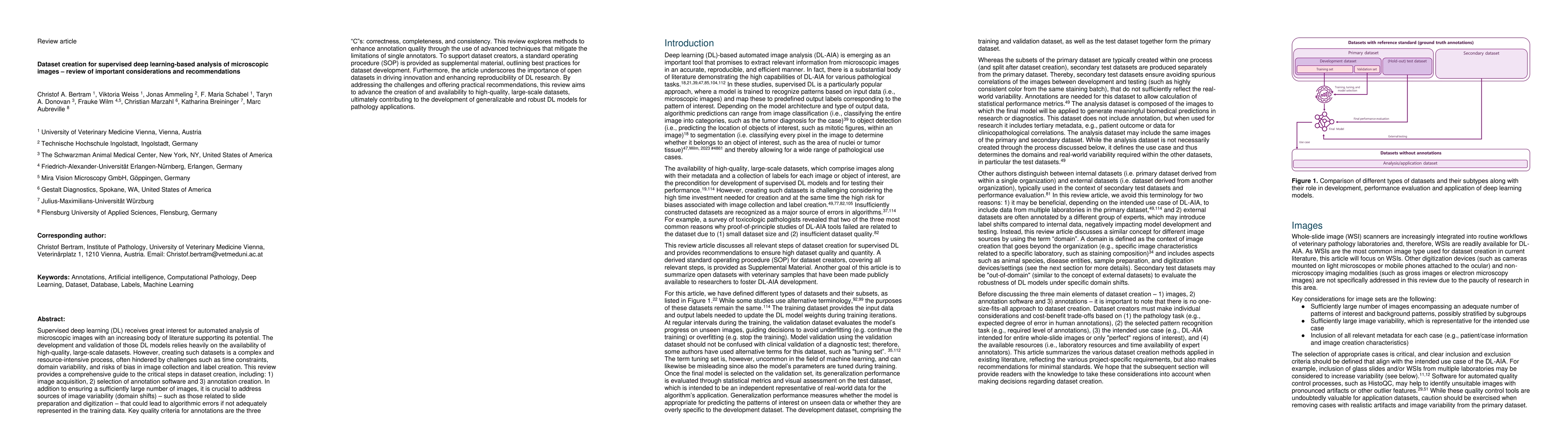
Supervised deep learning (DL) receives great interest for automated analysis of microscopic images with an increasing body of literature supporting its potential. The development and validation of tho...
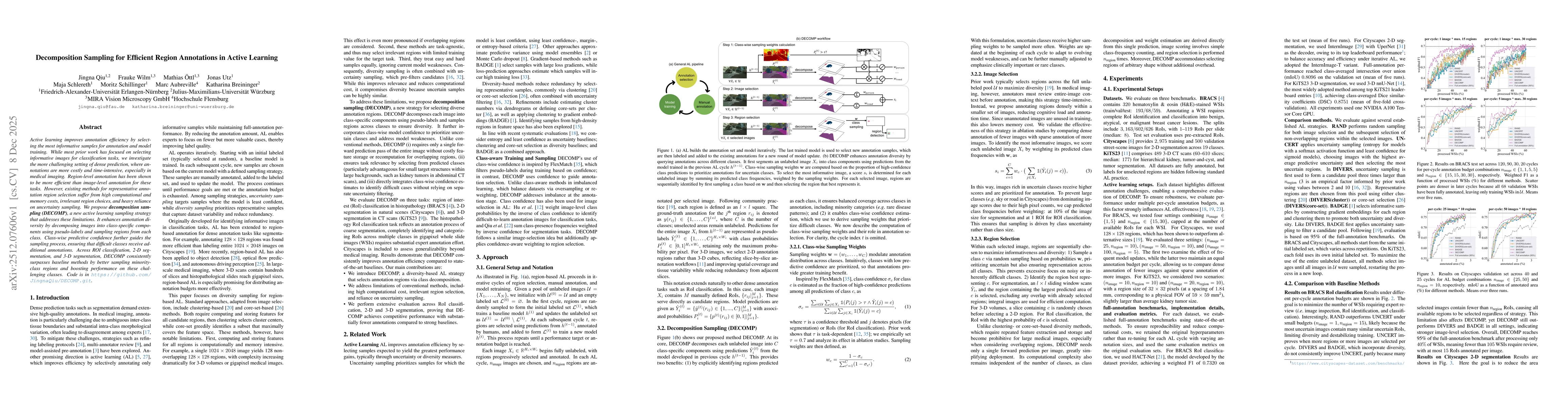
Active learning improves annotation efficiency by selecting the most informative samples for annotation and model training. While most prior work has focused on selecting informative images for classi...
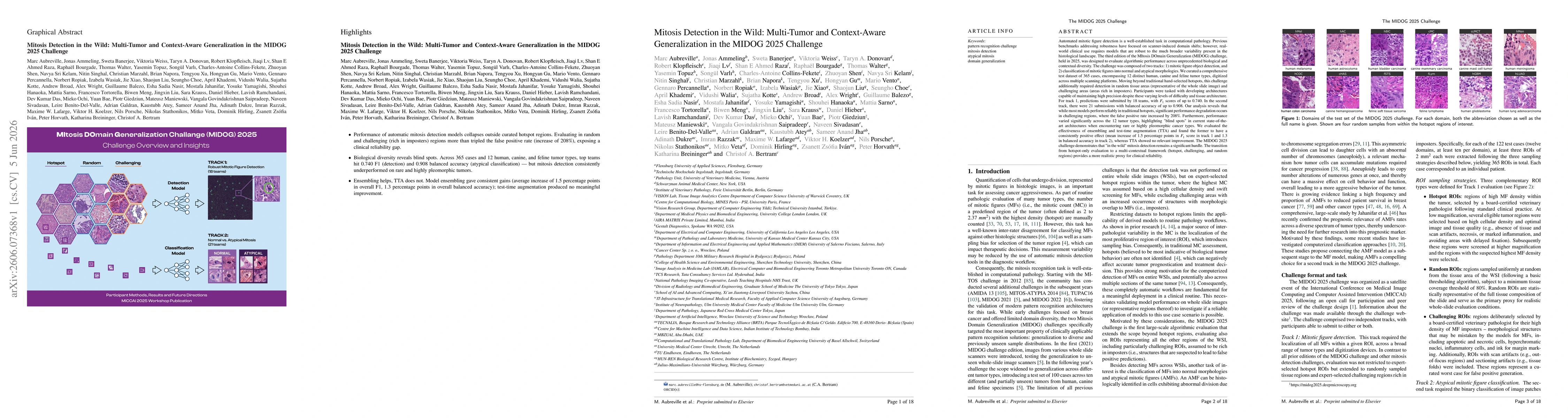
Automated mitosis detection is a well-established task in computational pathology. While previous benchmarks focused on scanner-induced domain shift, clinical "real-world" application requires models ...