Employing Graph Representations for Cell-level Characterization of Melanoma MELC Samples
Publication
Metrics
AI Quick Summary
This paper proposes a graph-based approach for characterizing melanoma samples using Multi-Epitope-Ligand Cartography (MELC) data, which trains a graph neural network to achieve a classification accuracy of 87%, outperforming existing methods by 10%.
Paper Preview
Abstract
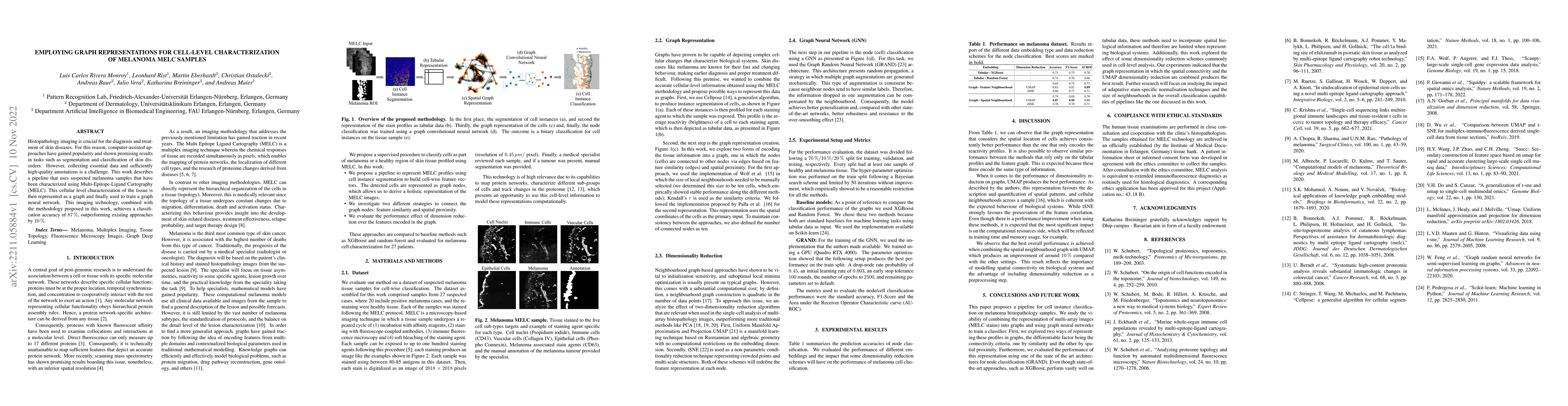
Histopathology imaging is crucial for the diagnosis and treatment of skin diseases. For this reason, computer-assisted approaches have gained popularity and shown promising results in tasks such as segmentation and classification of skin disorders. However, collecting essential data and sufficiently high-quality annotations is a challenge. This work describes a pipeline that uses suspected melanoma samples that have been characterized using Multi-Epitope-Ligand Cartography (MELC). This cellular-level tissue characterisation is then represented as a graph and used to train a graph neural network. This imaging technology, combined with the methodology proposed in this work, achieves a classification accuracy of 87%, outperforming existing approaches by 10%.
AI Key Findings
Get AI-generated insights about this paper's methodology, results, significance, and more — seven facets brought into focus.
Impact
Paper Details
Authors
PDF Preview
Key Terms
Citation Network
Current paper (gray), citations (green), references (blue)
Display is limited for performance on very large graphs.
Discussion 0