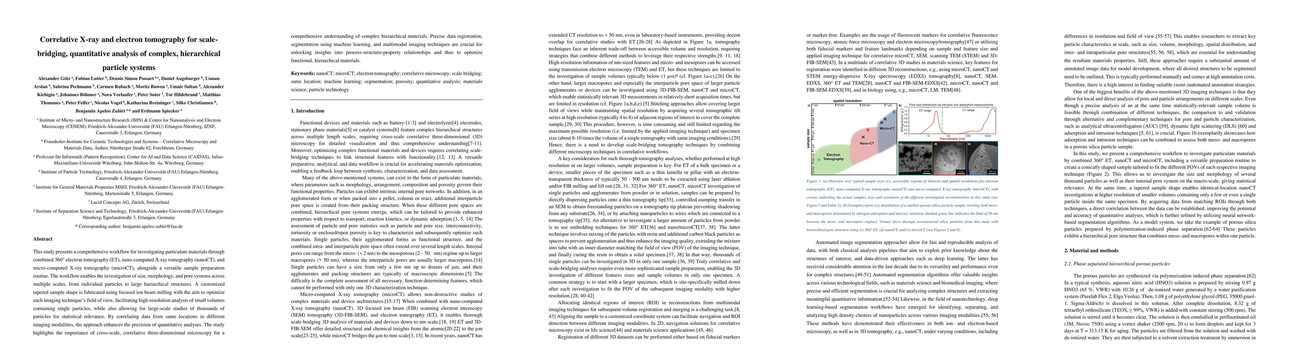
This study presents a comprehensive workflow for investigating particulate
materials through combined 360{\deg} electron tomography (ET), nano-computed
X-ray tomography (nanoCT), and micro-computed X-ray tomography (microCT),
alongside a versatile sample preparation routine. The workflow enables the
investigation of size, morphology, and pore systems across multiple scales,
from individual particles to large hierarchical structures. A customized
tapered sample shape is fabricated using focused ion beam milling with the aim
to optimize each imaging technique's field of view, facilitating
high-resolution analysis of small volumes containing single particles, while
also allowing for large-scale studies of thousands of particles for statistical
relevance. By correlating data from same locations in different imaging
modalities, the approach enhances the precision of quantitative analyses. The
study highlights the importance of cross-scale, correlative three-dimensional
microscopy for a comprehensive understanding of complex hierarchical materials.
Precise data registration, segmentation using machine learning, and multimodal
imaging techniques are crucial for unlocking insights into
process-structure-property relationships and thus to optimize functional,
hierarchical materials.
Discussion 0