Automatic 3D Ultrasound Segmentation of Uterus Using Deep Learning
Publication
Metrics
AI Quick Summary
This paper develops automatic 3D ultrasound segmentation of the uterus using deep learning to eliminate the need for manual initialization in semi-automatic algorithms. The study employs 2D UNet-based networks trained on individual planes and a comprehensive 3D volume approach to achieve precise uterine boundary delineation.
Paper Preview
Abstract
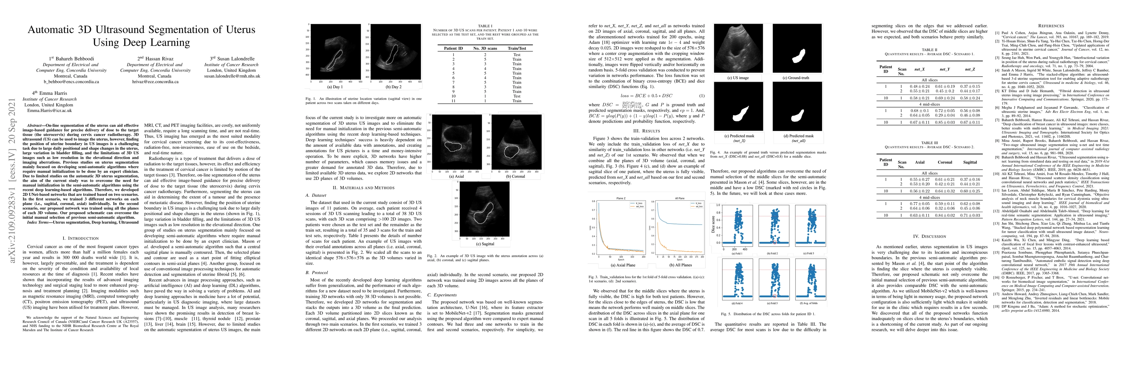
On-line segmentation of the uterus can aid effective image-based guidance for precise delivery of dose to the target tissue (the uterocervix) during cervix cancer radiotherapy. 3D ultrasound (US) can be used to image the uterus, however, finding the position of uterine boundary in US images is a challenging task due to large daily positional and shape changes in the uterus, large variation in bladder filling, and the limitations of 3D US images such as low resolution in the elevational direction and imaging aberrations. Previous studies on uterus segmentation mainly focused on developing semi-automatic algorithms where require manual initialization to be done by an expert clinician. Due to limited studies on the automatic 3D uterus segmentation, the aim of the current study was to overcome the need for manual initialization in the semi-automatic algorithms using the recent deep learning-based algorithms. Therefore, we developed 2D UNet-based networks that are trained based on two scenarios. In the first scenario, we trained 3 different networks on each plane (i.e., sagittal, coronal, axial) individually. In the second scenario, our proposed network was trained using all the planes of each 3D volume. Our proposed schematic can overcome the initial manual selection of previous semi-automatic algorithm.
AI Key Findings
Get AI-generated insights about this paper's methodology, results, significance, and more — seven facets brought into focus.
Impact
Paper Details
Authors
PDF Preview
Key Terms
Citation Network
Current paper (gray), citations (green), references (blue)
Display is limited for performance on very large graphs.
Discussion 0