Automatic ultrasound vessel segmentation with deep spatiotemporal context learning
Publication
Metrics
AI Quick Summary
This paper proposes deep learning methods for real-time segmentation of small vessels in ultrasound images, leveraging spatiotemporal context to improve accuracy. The approach combines temporal, spatial, and feature-aware embeddings, and outperforms baseline methods when evaluated on femoral and tibial artery scans.
Paper Preview
Abstract
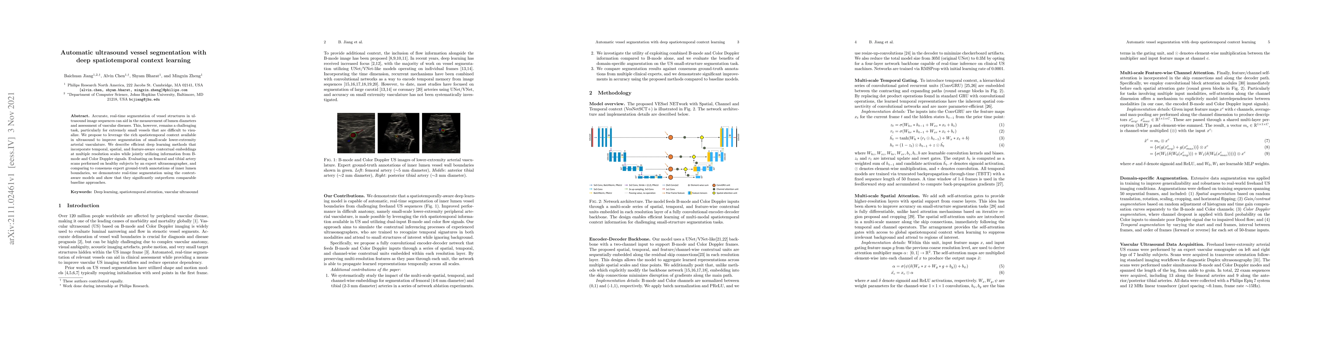
Accurate, real-time segmentation of vessel structures in ultrasound image sequences can aid in the measurement of lumen diameters and assessment of vascular diseases. This, however, remains a challenging task, particularly for extremely small vessels that are difficult to visualize. We propose to leverage the rich spatiotemporal context available in ultrasound to improve segmentation of small-scale lower-extremity arterial vasculature. We describe efficient deep learning methods that incorporate temporal, spatial, and feature-aware contextual embeddings at multiple resolution scales while jointly utilizing information from B-mode and Color Doppler signals. Evaluating on femoral and tibial artery scans performed on healthy subjects by an expert ultrasonographer, and comparing to consensus expert ground-truth annotations of inner lumen boundaries, we demonstrate real-time segmentation using the context-aware models and show that they significantly outperform comparable baseline approaches.
AI Key Findings
Get AI-generated insights about this paper's methodology, results, significance, and more — seven facets brought into focus.
Impact
Paper Details
Authors
PDF Preview
Key Terms
Citation Network
Current paper (gray), citations (green), references (blue)
Display is limited for performance on very large graphs.
Discussion 0