Automatic Cerebral Vessel Extraction in TOF-MRA Using Deep Learning
Publication
Metrics
AI Quick Summary
This study explores deep learning methods for automatic cerebral vessel segmentation in Time-of-Flight Magnetic Resonance Angiographs (TOF-MRAs), finding that 2D and 3D U-Nets with data augmentation techniques like Gaussian blur, rotation, and flipping yield the best results, achieving Dice Similarity Coefficient scores ranging from 0.72 to 0.83.
Paper Preview
Abstract
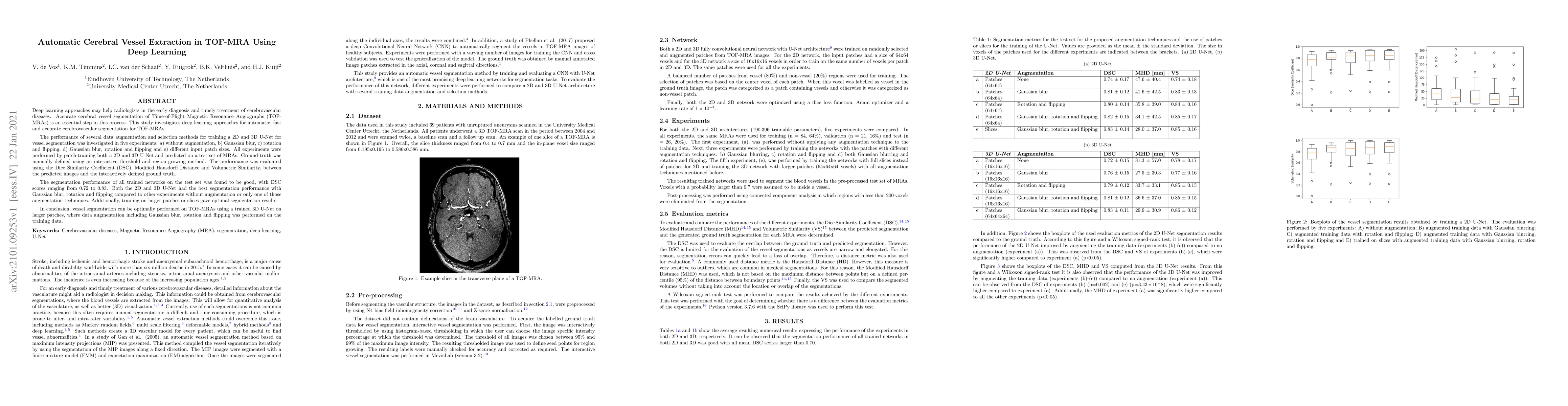
Deep learning approaches may help radiologists in the early diagnosis and timely treatment of cerebrovascular diseases. Accurate cerebral vessel segmentation of Time-of-Flight Magnetic Resonance Angiographs (TOF-MRAs) is an essential step in this process. This study investigates deep learning approaches for automatic, fast and accurate cerebrovascular segmentation for TOF-MRAs. The performance of several data augmentation and selection methods for training a 2D and 3D U-Net for vessel segmentation was investigated in five experiments: a) without augmentation, b) Gaussian blur, c) rotation and flipping, d) Gaussian blur, rotation and flipping and e) different input patch sizes. All experiments were performed by patch-training both a 2D and 3D U-Net and predicted on a test set of MRAs. Ground truth was manually defined using an interactive threshold and region growing method. The performance was evaluated using the Dice Similarity Coefficient (DSC), Modified Hausdorff Distance and Volumetric Similarity, between the predicted images and the interactively defined ground truth. The segmentation performance of all trained networks on the test set was found to be good, with DSC scores ranging from 0.72 to 0.83. Both the 2D and 3D U-Net had the best segmentation performance with Gaussian blur, rotation and flipping compared to other experiments without augmentation or only one of those augmentation techniques. Additionally, training on larger patches or slices gave optimal segmentation results. In conclusion, vessel segmentation can be optimally performed on TOF-MRAs using a trained 3D U-Net on larger patches, where data augmentation including Gaussian blur, rotation and flipping was performed on the training data.
AI Key Findings
Get AI-generated insights about this paper's methodology, results, significance, and more — seven facets brought into focus.
Impact
Paper Details
Authors
PDF Preview
Key Terms
Citation Network
Current paper (gray), citations (green), references (blue)
Display is limited for performance on very large graphs.
Discussion 0