Deep Learning-based Segmentation of Cerebral Aneurysms in 3D TOF-MRA using Coarse-to-Fine Framework
Publication
Metrics
AI Quick Summary
This paper proposes a deep learning-based segmentation framework for cerebral aneurysms in 3D TOF-MRA using a coarse-to-fine approach, achieving superior accuracy compared to existing methods. The framework combines DeepMedic for coarse segmentation and a dual-channel SE_3D U-Net for fine segmentation, demonstrating high DSC and low HD on validation and test cohorts.
Paper Preview
Abstract
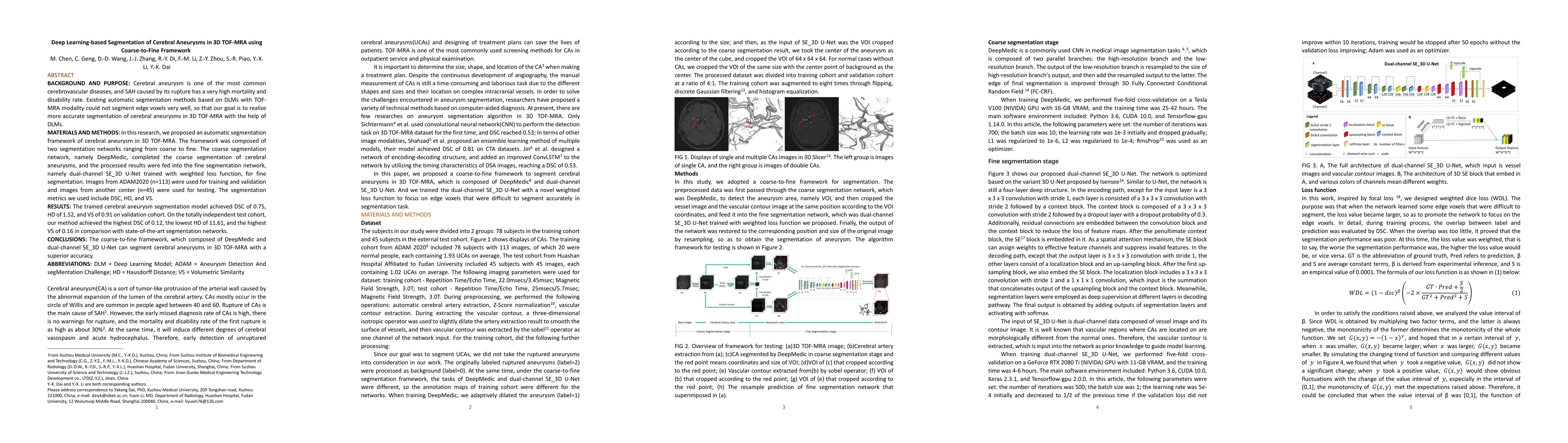
BACKGROUND AND PURPOSE: Cerebral aneurysm is one of the most common cerebrovascular diseases, and SAH caused by its rupture has a very high mortality and disability rate. Existing automatic segmentation methods based on DLMs with TOF-MRA modality could not segment edge voxels very well, so that our goal is to realize more accurate segmentation of cerebral aneurysms in 3D TOF-MRA with the help of DLMs. MATERIALS AND METHODS: In this research, we proposed an automatic segmentation framework of cerebral aneurysm in 3D TOF-MRA. The framework was composed of two segmentation networks ranging from coarse to fine. The coarse segmentation network, namely DeepMedic, completed the coarse segmentation of cerebral aneurysms, and the processed results were fed into the fine segmentation network, namely dual-channel SE_3D U-Net trained with weighted loss function, for fine segmentation. Images from ADAM2020 (n=113) were used for training and validation and images from another center (n=45) were used for testing. The segmentation metrics we used include DSC, HD, and VS. RESULTS: The trained cerebral aneurysm segmentation model achieved DSC of 0.75, HD of 1.52, and VS of 0.91 on validation cohort. On the totally independent test cohort, our method achieved the highest DSC of 0.12, the lowest HD of 11.61, and the highest VS of 0.16 in comparison with state-of-the-art segmentation networks. CONCLUSIONS: The coarse-to-fine framework, which composed of DeepMedic and dual-channel SE_3D U-Net can segment cerebral aneurysms in 3D TOF-MRA with a superior accuracy.
AI Key Findings
Get AI-generated insights about this paper's methodology, results, significance, and more — seven facets brought into focus.
Impact
Paper Details
Authors
PDF Preview
Key Terms
Citation Network
Current paper (gray), citations (green), references (blue)
Display is limited for performance on very large graphs.
Discussion 0