Neoadjuvant chemotherapy (NAC) has become a standard clinical practice for
tumor downsizing in breast cancer with 18F-FDG Positron Emission Tomography
(PET). Our work aims to leverage PET imaging for the segmentation of breast
lesions. The focus is on developing an automated system that accurately
segments primary tumor regions and extracts key biomarkers from these areas to
provide insights into the evolution of breast cancer following the first course
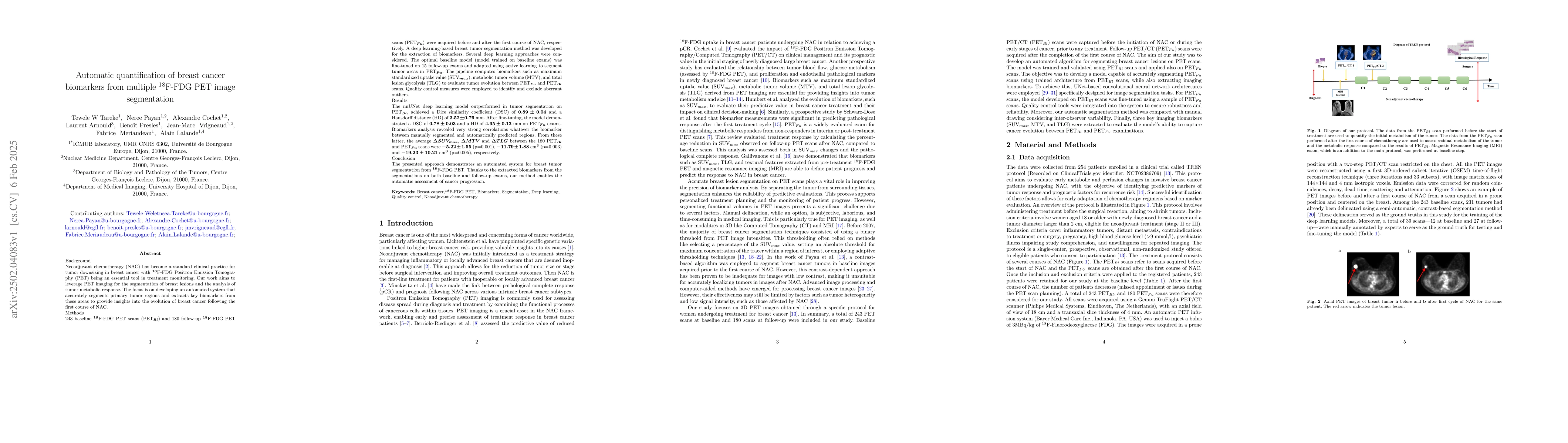
of NAC. 243 baseline 18F-FDG PET scans (PET_Bl) and 180 follow-up 18F-FDG PET
scans (PET_Fu) were acquired before and after the first course of NAC,
respectively. Firstly, a deep learning-based breast tumor segmentation method
was developed. The optimal baseline model (model trained on baseline exams) was
fine-tuned on 15 follow-up exams and adapted using active learning to segment
tumor areas in PET_Fu. The pipeline computes biomarkers such as maximum
standardized uptake value (SUVmax), metabolic tumor volume (MTV), and total
lesion glycolysis (TLG) to evaluate tumor evolution between PET_Fu and PET_Bl.
Quality control measures were employed to exclude aberrant outliers. The nnUNet
deep learning model outperformed in tumor segmentation on PET_Bl, achieved a
Dice similarity coefficient (DSC) of 0.89 and a Hausdorff distance (HD) of 3.52
mm. After fine-tuning, the model demonstrated a DSC of 0.78 and a HD of 4.95 mm
on PET_Fu exams. Biomarkers analysis revealed very strong correlations whatever
the biomarker between manually segmented and automatically predicted regions.
The significant average decrease of SUVmax, MTV and TLG were 5.22, 11.79 cm3
and 19.23 cm3, respectively. The presented approach demonstrates an automated
system for breast tumor segmentation from 18F-FDG PET. Thanks to the extracted
biomarkers, our method enables the automatic assessment of cancer progression.
Discussion 0