Automatic segmentation of skin lesions using deep learning
Publication
Metrics
AI Quick Summary
Researchers developed a fully automated method to accurately segment skin lesion boundaries from dermoscopic images using a U-net deep learning network. Their approach combined pre-processing steps with intensity, color, and texture enhancement, as well as post-processing steps including morphological operations and contour identification.
Paper Preview
Abstract
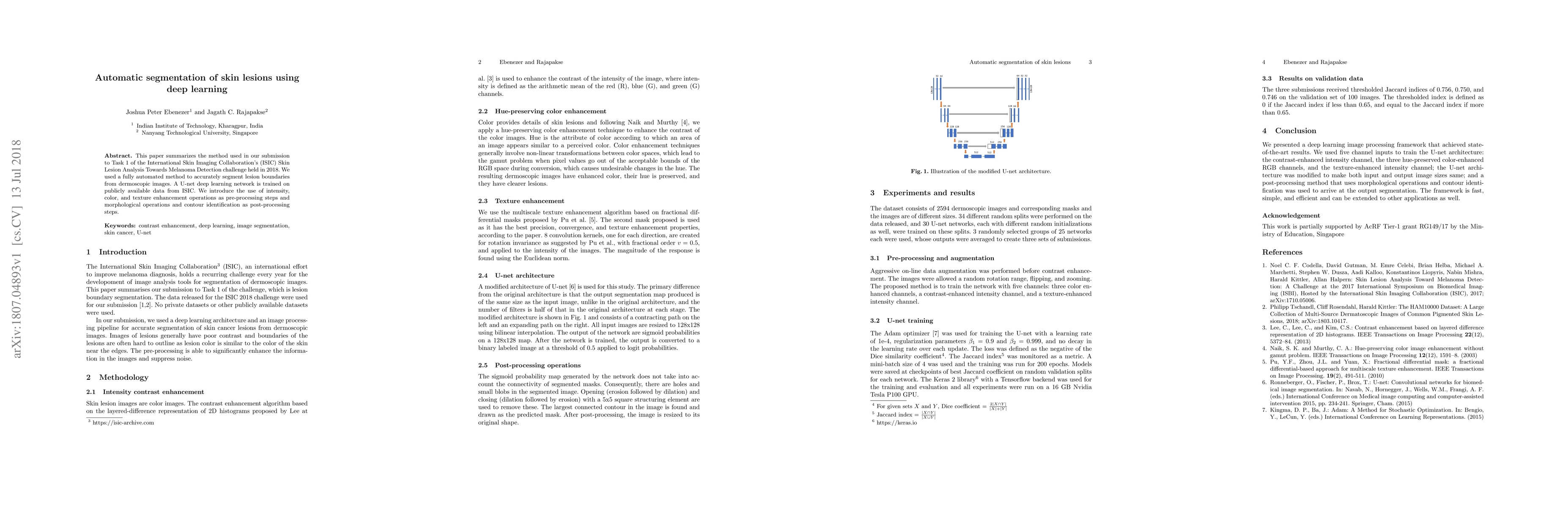
This paper summarizes the method used in our submission to Task 1 of the International Skin Imaging Collaboration's (ISIC) Skin Lesion Analysis Towards Melanoma Detection challenge held in 2018. We used a fully automated method to accurately segment lesion boundaries from dermoscopic images. A U-net deep learning network is trained on publicly available data from ISIC. We introduce the use of intensity, color, and texture enhancement operations as pre-processing steps and morphological operations and contour identification as post-processing steps.
AI Key Findings
Get AI-generated insights about this paper's methodology, results, significance, and more — seven facets brought into focus.
Impact
Paper Details
PDF Preview
Key Terms
Citation Network
Current paper (gray), citations (green), references (blue)
Display is limited for performance on very large graphs.
Discussion 0