Publication
Metrics
AI Quick Summary
This paper proposes an axial super-resolution evanescent wave tomography (AxSET) method to overcome the axial diffraction limit in optical tomography, achieving axial super-resolution using conventional evanescent wave microscopes. The method, validated through simulations and experiments, reconstructs microtubules in HeLa cells with an axial resolution of approximately 130 nm without requiring additional components or sample preparation.
Paper Preview
Abstract
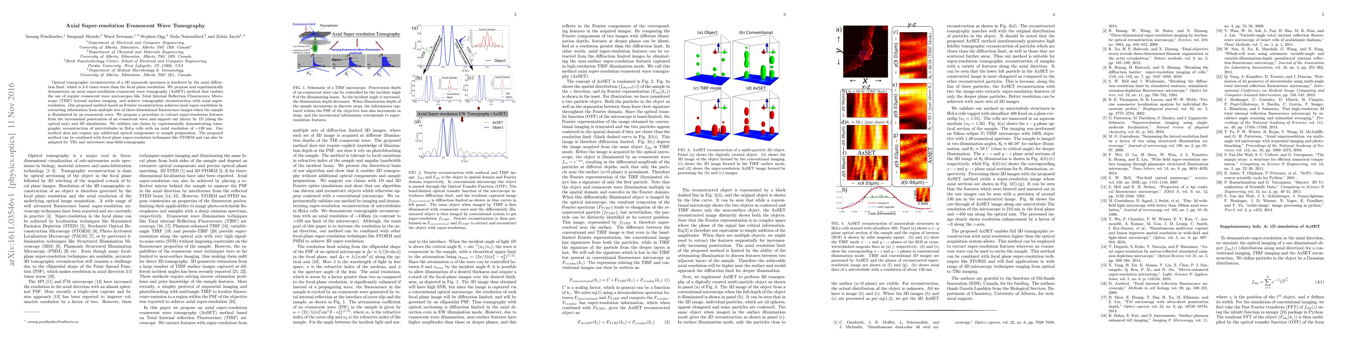
Optical tomographic reconstruction of a 3D nanoscale specimen is hindered by the axial diffraction limit, which is 2-3 times worse than the focal plane resolution. We propose and experimentally demonstrate an axial super-resolution evanescent wave tomography (AxSET) method that enables the use of regular evanescent wave microscopes like Total Internal Reflection Fluorescence Microscope (TIRF) beyond surface imaging, and achieve tomographic reconstruction with axial super-resolution. Our proposed method based on Fourier reconstruction achieves axial super-resolution by extracting information from multiple sets of three-dimensional fluorescence images when the sample is illuminated by an evanescent wave. We propose a procedure to extract super-resolution features from the incremental penetration of an evanescent wave and support our theory by 1D (along the optical axis) and 3D simulations. We validate our claims by experimentally demonstrating tomographic reconstruction of microtubules in HeLa cells with an axial resolution of $\sim$130 nm. Our method does not require any additional optical components or sample preparation. The proposed method can be combined with focal plane super-resolution techniques like STORM and can also be adapted for THz and microwave near-field tomography.
AI Key Findings
Get AI-generated insights about this paper's methodology, results, significance, and more — seven facets brought into focus.
Impact
Paper Details
PDF Preview
Key Terms
Citation Network
Current paper (gray), citations (green), references (blue)
Display is limited for performance on very large graphs.
Discussion 0