Cardiopulmonary resuscitation (CPR) is one of the essential tools to ensure
oxygen supply during cardiac arrest. However, the precise effects of chest
compression are not quantifiable to this day. This often results in a low
quality of chest compressions even if performed by health-care professionals.
One solution could be provided by quantification of blood flow via ultrasonic
Doppler measurements, to guide first responders in their efforts. This paper
presents an approach to address the issue of limited time, anatomical know how
and limitations of system configuration during emergency scenarios. An approach
for automated vessel identification with three different phases was developed,
featuring a new sensor probe for ultrasonic measurements with non symmetrically
angled piezo ceramics. The probe was used with prototype ultrasound hardware in
a laboratory setup for Pulsed Wave Doppler (PW Doppler). In an initial
measurement a qualitative flow was approximated to examine valuable measurement
positions on a phantom. Afterwards an iterative mode was used for depth
depending frequency measurements with score calculation of flow periodicity and
signal power. The configuration with the best score was used for a prolonged
monitoring mode. Flow values were compared to data of an industrial
flow-sensor. Flow-sensor data showed an average coefficient of determination of
0.97 with an average root mean square error of 3.84 ml/s. With the proposed
hardware and software solutions a basis for future developments was made, which
could lead to a fully automated vessel identification during CPR. This device
could provide first responders as well as clinical staff with vital information
about CPR efficiency that has yet to be included into the therapy of people
during cardiac arrest.
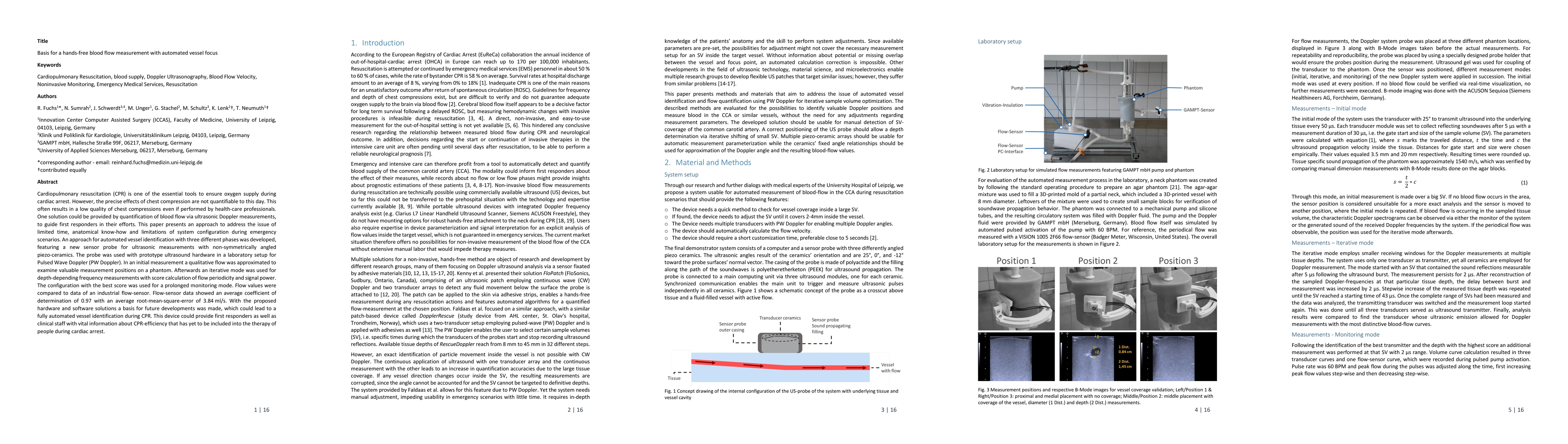
Discussion 0