Blood vessel segmentation in en-face OCTA images: a frequency based method
Publication
Metrics
AI Quick Summary
This paper introduces a novel frequency-based segmentation method using Gabor filter banks for blood vessel analysis in en-face OCTA images. The method is evaluated both qualitatively and quantitatively, showing good agreement with expert visual assessments and automated device values, and suggesting adaptive local vessel density maps for detailed retinal blood flow analysis.
Paper Preview
Abstract
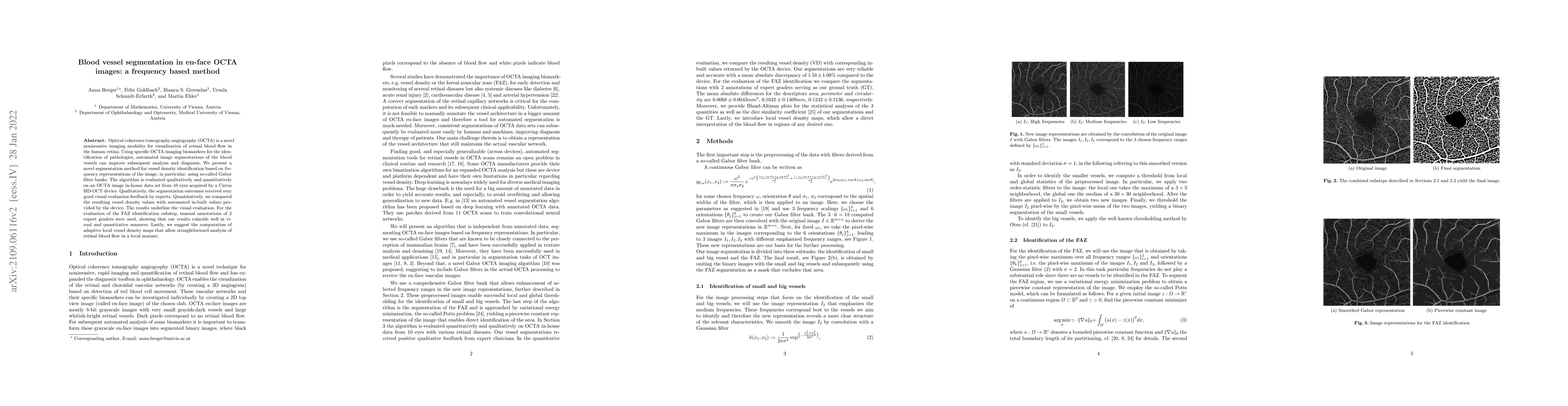
Optical coherence tomography angiography (OCTA) is a novel noninvasive imaging modality for visualization of retinal blood flow in the human retina. Using specific OCTA imaging biomarkers for the identification of pathologies, automated image segmentations of the blood vessels can improve subsequent analysis and diagnosis. We present a novel segmentation method for vessel density identification based on frequency representations of the image, in particular, using so-called Gabor filter banks. The algorithm is evaluated qualitatively and quantitatively on an OCTA image in-house data set from $10$ eyes acquired by a Cirrus HD-OCT device. Qualitatively, the segmentation outcomes received very good visual evaluation feedback by experts. Quantitatively, we compared the resulting vessel density values with automated in-built values provided by the device. The results underline the visual evaluation. For the evaluation of the FAZ identification substep, manual annotations of $2$ expert graders were used, showing that our results coincide well in visual and quantitative manners. Lastly, we suggest the computation of adaptive local vessel density maps that allow straightforward analysis of retinal blood flow in a local manner.
AI Key Findings
Get AI-generated insights about this paper's methodology, results, significance, and more — seven facets brought into focus.
Impact
Paper Details
Authors
PDF Preview
Key Terms
Citation Network
Current paper (gray), citations (green), references (blue)
Display is limited for performance on very large graphs.
Discussion 0