Beam Cross Sections Create Mixtures: Improving Feature Localization in Secondary Electron Imaging
Publication
Metrics
Paper Preview
Abstract
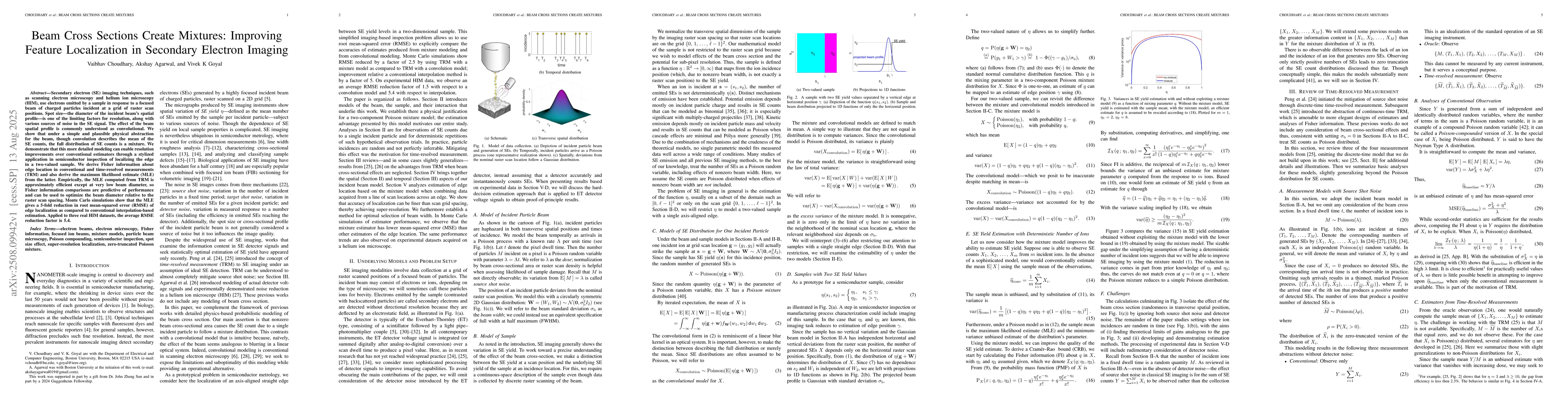
Secondary electron (SE) imaging techniques, such as scanning electron microscopy and helium ion microscopy (HIM), use electrons emitted by a sample in response to a focused beam of charged particles incident at a grid of raster scan positions. Spot size -- the diameter of the incident beam's spatial profile -- is one of the limiting factors for resolution, along with various sources of noise in the SE signal. The effect of the beam spatial profile is commonly understood as convolutional. We show that under a simple and plausible physical abstraction for the beam, though convolution describes the mean of the SE counts, the full distribution of SE counts is a mixture. We demonstrate that this more detailed modeling can enable resolution improvements over conventional estimators through a stylized application in semiconductor inspection of localizing the edge in a two-valued sample. We derive Fisher information about edge location in conventional and time-resolved measurements (TRM) and also derive the maximum likelihood estimate (MLE) from the latter. Empirically, the MLE computed from TRM is approximately efficient except at very low beam diameter, so Fisher information comparisons are predictive of performance and can be used to optimize the beam diameter relative to the raster scan spacing. Monte Carlo simulations show that the MLE gives a 5-fold reduction in root mean-squared error (RMSE) of edge localization as compared to conventional interpolation-based estimation. Applied to three real HIM datasets, the average RMSE reduction factor is 5.4.
AI Key Findings
Get AI-generated insights about this paper's methodology, results, significance, and more — seven facets brought into focus.
Discussion 0