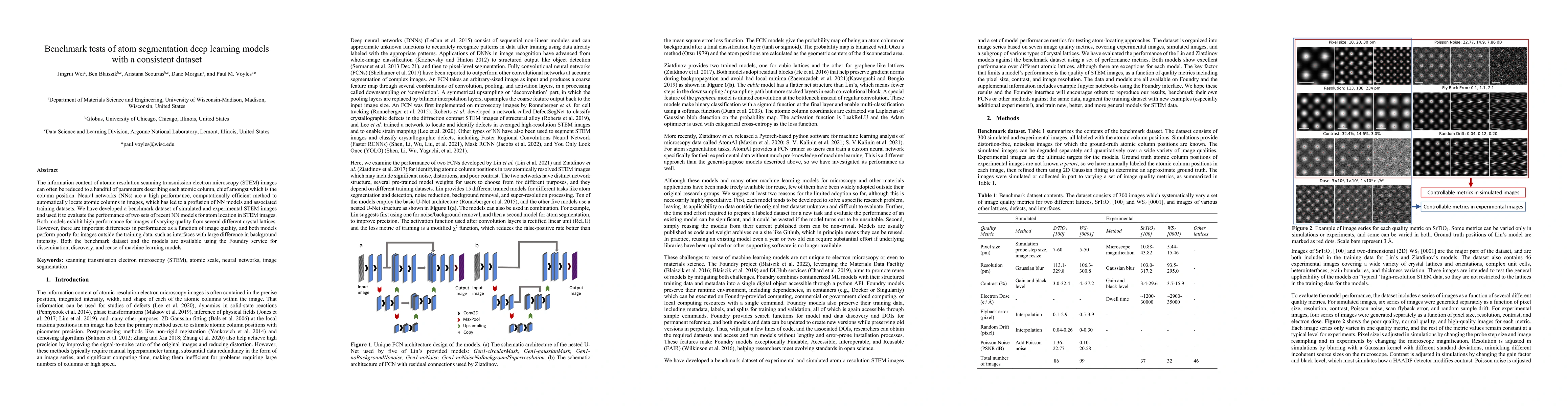
This paper benchmarks deep learning models for atom segmentation in STEM images using a consistent dataset, finding that while the models perform well on training data, they struggle with images outside the training scope, especially those with significant background intensity differences. The benchmark dataset and models are made available for dissemination via the Foundry service.
The information content of atomic resolution scanning transmission electron microscopy (STEM) images can often be reduced to a handful of parameters describing each atomic column, chief amongst which is the column position. Neural networks (NNs) are a high performance, computationally efficient method to automatically locate atomic columns in images, which has led to a profusion of NN models and associated training datasets. We have developed a benchmark dataset of simulated and experimental STEM images and used it to evaluate the performance of two sets of recent NN models for atom location in STEM images. Both models exhibit high performance for images of varying quality from several different crystal lattices. However, there are important differences in performance as a function of image quality, and both models perform poorly for images outside the training data, such as interfaces with large difference in background intensity. Both the benchmark dataset and the models are available using the Foundry service for dissemination, discovery, and reuse of machine learning models.
Get AI-generated insights about this paper's methodology, results, and significance.
Current paper (gray), citations (green), references (blue)
Chao Tao, Haiyang Wu, Dandan Zhong et al.
Haibo Wang, Lutfu Sua, Jun Huang
Ujjwal Verma, Srinivasan Murali, Jaya Sreevalsan-Nair et al.
Fabio Remondino, Xiaowei Yu, Joel Kostensalo et al.
Guan Gui, Zeeshan Kaleem, Misha Urooj Khan et al.
Title | Authors | Year | Actions |
---|---|---|---|
Predicting Performance of Object Detection Models in Electron Microscopy Using Random Forests
|
Ni Li, Ryan Jacobs, Matthew J. Lynch, Vidit Agrawal, Kevin G. Field, Dane Morgan
|
2025
|
View Paper |
Accurate quantification of dislocation loops in complex functional alloys enabled by deep learning image analysis
|
Thomas Bilyk, A. Goryaeva, M. Marinica, Camille Flament, Catherine Sabathier, Eric Leroy, Marie Loyer-Prost, Estelle Meslin
|
2024
|
View Paper |
Localization and segmentation of atomic columns in supported nanoparticles for fast scanning transmission electron microscopy
|
Henrik Eliasson, Rolf Erni
|
2024
|
View Paper |
Generalization Across Experimental Parameters in Neural Network Analysis of High-Resolution Transmission Electron Microscopy Datasets.
|
Katherine Sytwu, L. Rangel DaCosta, Mary C Scott
|
2024
|
View Paper |
JARVIS-Leaderboard: a large scale benchmark of materials design methods
|
K. Choudhary, D. Wines, Kangming Li, K. Garrity, Vishu Gupta, A. Romero, J. Krogel, Kayahan Saritas, Addis S. Fuhr, P. Ganesh, P. Kent, Keqiang Yan, Yu-Ching Lin, Shuiwang Ji, B. Blaiszik, Patrick Reiser, Pascal Friederich, Ankit Agrawal, P. Tiwary, E. Beyerle, Peter Minch, T. Rhone, I. Takeuchi, R. Wexler, Arun Mannodi-Kanakkithodi, E. Ertekin, Avanish Mishra, Nithin Mathew, Sterling G. Baird, M. Wood, Andrew Rohskopf, J. Hattrick-Simpers, Shih-Han Wang, Luke E. K. Achenie, Hongliang Xin, M. Williams, A. Biacchi, F. Tavazza
|
2023
|
View Paper |
Generalization Across Experimental Parameters in Machine Learning Analysis of High Resolution Transmission Electron Microscopy Datasets
|
Katherine Sytwu, L. Dacosta, M. Scott
|
2023
|
View Paper |
Automatic identification of crystal structures and interfaces via artificial-intelligence-based electron microscopy
|
Andreas Leitherer, Byung Chul Yeo, C. Liebscher, L. Ghiringhelli
|
2023
|
View Paper |
Comments (0)