Publication
Metrics
AI Quick Summary
This paper investigates the integration of intravascular optical coherence tomography (IVOCT) and magnetic particle imaging (MPI) for bimodal intravascular volumetric imaging. The study establishes a method to estimate the IVOCT pullback path using 3D MPI data, achieving accurate reconstruction of vessel shapes by combining the high-resolution imaging of IVOCT with the spatial orientation provided by MPI.
Paper Preview
Abstract
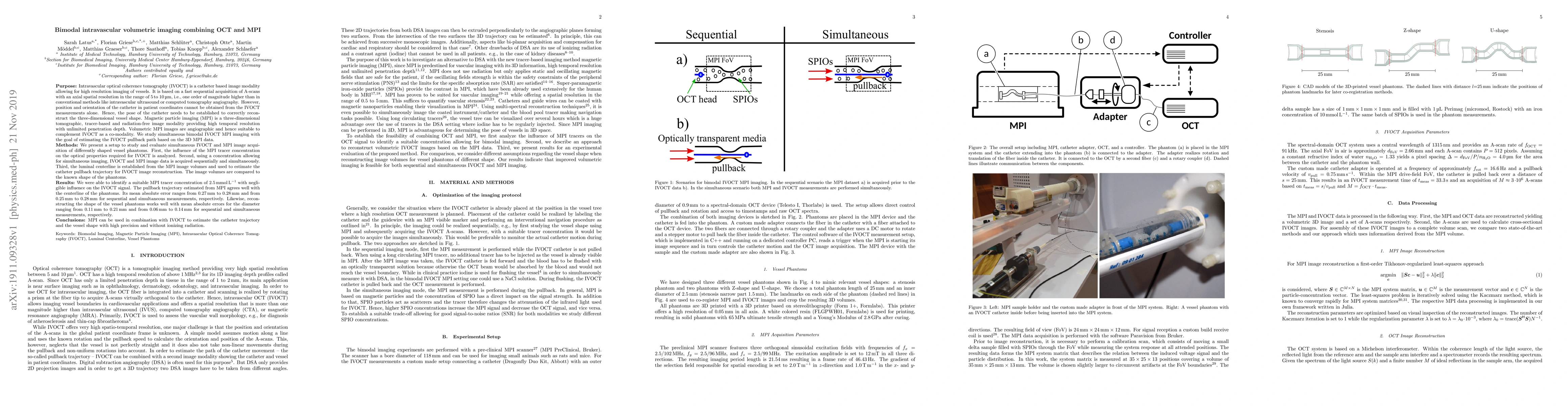
Intravascular optical coherence tomography (IVOCT) is a catheter based image modality allowing for high resolution imaging of vessels. It is based on a fast sequential acquisition of A-scans with an axial spatial resolution in the range of 5 to 10 {\mu}m, i.e., one order of magnitude higher than in conventional methods like intravascular ultrasound or computed tomography angiography. However, position and orientation of the catheter in patient coordinates cannot be obtained from the IVOCT measurements alone. Hence, the pose of the catheter needs to be established to correctly reconstruct the three-dimensional vessel shape. Magnetic particle imaging (MPI) is a three-dimensional tomographic, tracer-based and radiation-free image modality providing high temporal resolution with unlimited penetration depth. Volumetric MPI images are angiographic and hence suitable to complement IVOCT as a co-modality. We study simultaneous bimodal IVOCT MPI imaging with the goal of estimating the IVOCT pullback path based on the 3D MPI data. We present a setup to study and evaluate simultaneous IVOCT and MPI image acquisition of differently shaped vessel phantoms. First, the infuence of the MPI tracer concentration on the optical properties required for IVOCT is analyzed. Second, using a concentration allowing for simultaneous imaging, IVOCT and MPI image data is acquired sequentially and simultaneously. Third, the luminal centerline is established from the MPI image volumes and used to estimate the catheter pullback trajectory for IVOCT image reconstruction. The image volumes are compared to the known shape of the phantoms. We were able to identify a suitable MPI tracer concentration of 2.5 mmol/L with negligible influence on the IVOCT signal. The pullback trajectory estimated from MPI agrees well with the centerline of the phantoms. (...)
AI Key Findings
Get AI-generated insights about this paper's methodology, results, significance, and more — seven facets brought into focus.
Impact
Paper Details
Authors
PDF Preview
Key Terms
Citation Network
Current paper (gray), citations (green), references (blue)
Display is limited for performance on very large graphs.
Discussion 0