Quick Summary:

This paper proposes a universal digital filtering method for denoising volumetric retinal OCT and OCTA images, utilizing 3D shearlet transforms to effectively separate retinal tissue and vessels from noise. The 3D shearlet approach outperforms existing methods and demonstrates superior results compared to 2D filtering techniques.

Summary

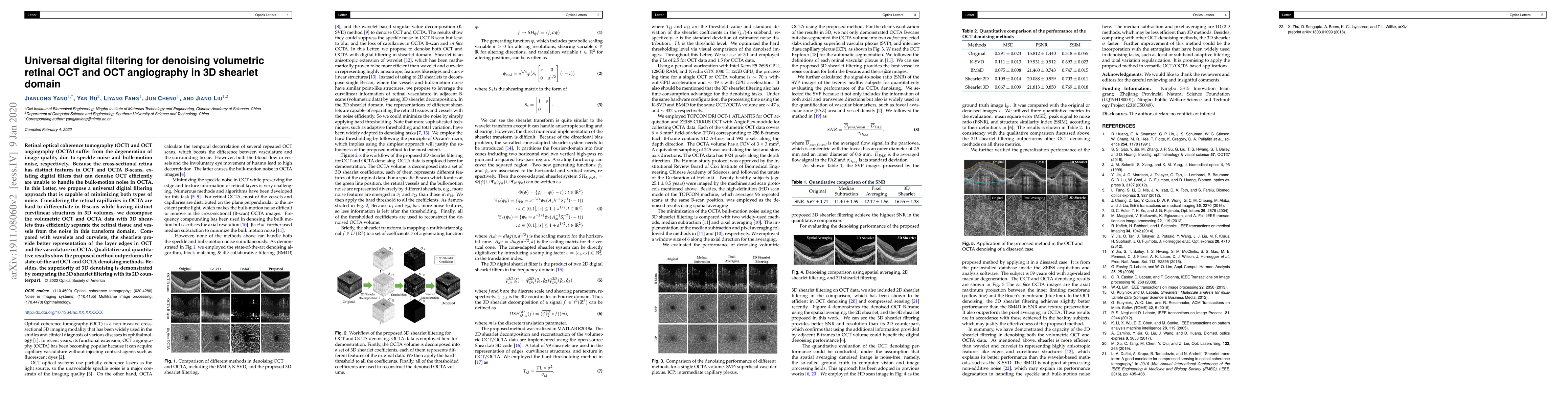
Retinal optical coherence tomography (OCT) and OCT angiography (OCTA) suffer from the degeneration of image quality due to speckle noise and bulk-motion noise, respectively. Because the cross-sectional retina has distinct features in OCT and OCTA B-scans, existing digital filters that can denoise OCT efficiently are unable to handle the bulk-motion noise in OCTA. In this Letter, we propose a universal digital filtering approach that is capable of minimizing both types of noise. Considering the retinal capillaries in OCTA are hard to differentiate in B-scans while having distinct curvilinear structures in 3D volumes, we decompose the volumetric OCT and OCTA data with 3D shearlets thus efficiently separate the retinal tissue and vessels from the noise in this transform domain. Compared with wavelets and curvelets, the shearlets provide better representation of the layer edges in OCT and the vasculature in OCTA. Qualitative and quantitative results show the proposed method outperforms the state-of-the-art OCT and OCTA denoising methods. Besides, the superiority of 3D denoising is demonstrated by comparing the 3D shearlet filtering with its 2D counterpart.

AI Key Findings

Get AI-generated insights about this paper's methodology, results, and significance.

Paper Details

PDF Preview

Download PDF

Key Terms

noise (0.260)
denoising (0.259)
filtering (0.234)
digital (0.210)
efficiently (0.197)
bulk (0.196)
distinct (0.190)
motion (0.170)
volumes (0.158)
method outperforms (0.151)
filters (0.147)
tomography (0.146)
outperforms state (0.144)
superiority (0.139)
counterpart (0.139)
letter (0.138)
minimizing (0.137)
suffer (0.136)
handle (0.134)
coherence (0.132)

Citation Network

Current paper (gray), citations (green), references (blue)

Current Paper
Citations
References
Click to view
Title Authors Year Actions
Dynamic Change of Amplitude for OCT Functional Imaging
Jianlong Yang, Haoran Zhang, Liu Chang, Chengfu Gu
2023
View Paper
Optical coherence tomography image despeckling based on tensor singular value decomposition and fractional edge detection
Ying Fang, Xiao Shao, Bangquan Liu, Hongli Lv
2023
View Paper
An optimized seed point selection method assists digital image correlation for measuring deformation from OCT images of fundus diseases
Xiaochuan Zhang, Zhiqing Li, Anqi Wang, Qieni Lu, Jingcheng Ye
2023
View Paper
Optical coherence tomography image despeckling based on edge feature-guided higher-order singular value decomposition
Hongli Lv
2023
View Paper
Self-supervised patient-specific features learning for OCT image classification
Leyuan Fang, Jiahuan Guo, Xingxin He, Muxing Li
2022
View Paper
Noise reduction by adaptive-SIN filtering for retinal OCT images
Yan Hu, Jianfeng Ren, Jianlong Yang, Ruibin Bai, Jiang Liu
2021
View Paper
A nonlocal deep image prior model to restore optical coherence tomographic images from gamma distributed speckle noise
A. Smitha, P. Jidesh
2021
View Paper
OCTA-500: A retinal dataset for optical coherence tomography angiography study
Mingchao Li, Kun Huang, Qiuzhuo Xu, Jiadong Yang, Yuhan Zhang, Zexuan Ji, Keren Xie, Songtao Yuan, Qinghuai Liu, Qiang Chen
2020
View Paper
Using a dual-tree complex wavelet transform for denoising an optical coherence tomography angiography blood vessel image
Huazong Liu, Shenghao Lin, Ye Chong, Dan Yu, Qin Jia, An Lin
2020
View Paper
基于光场幅度动态变化的OCT功能成像原理和应用(特邀)
杨建龙 Yang Jianlong, 张浩然 Zhang Haoran, 刘畅 Liu Chang, 谷成富 Gu Chengfu
2024
View Paper
Self-supervised Denoising and Bulk Motion Artifact Removal of 3D Optical Coherence Tomography Angiography of Awake Brain
Zhenghong Li, Jiaxiang Ren, Zhilin Zou, Kalyan Garigapati, C. Du, Yingtian Pan, Haibin Ling
2024
View Paper
Fusion denoising algorithm of optical coherence tomography image based on point-estimated and block-estimated
Huaiguang Chen
2021
View Paper

Comments (0)

This website uses cookies to ensure you get the best experience. Learn more