This study proposes a multi-atlas guided 3D fully convolutional network ensemble model (M-FCN) for segmenting brain regions from MRIs, addressing limitations of fixed-size patch models by using adaptive patch sizes and incorporating multi-atlas guidance for robust segmentation. The method demonstrated superior performance compared to existing models in segmenting both subcortical and larger brain structures.
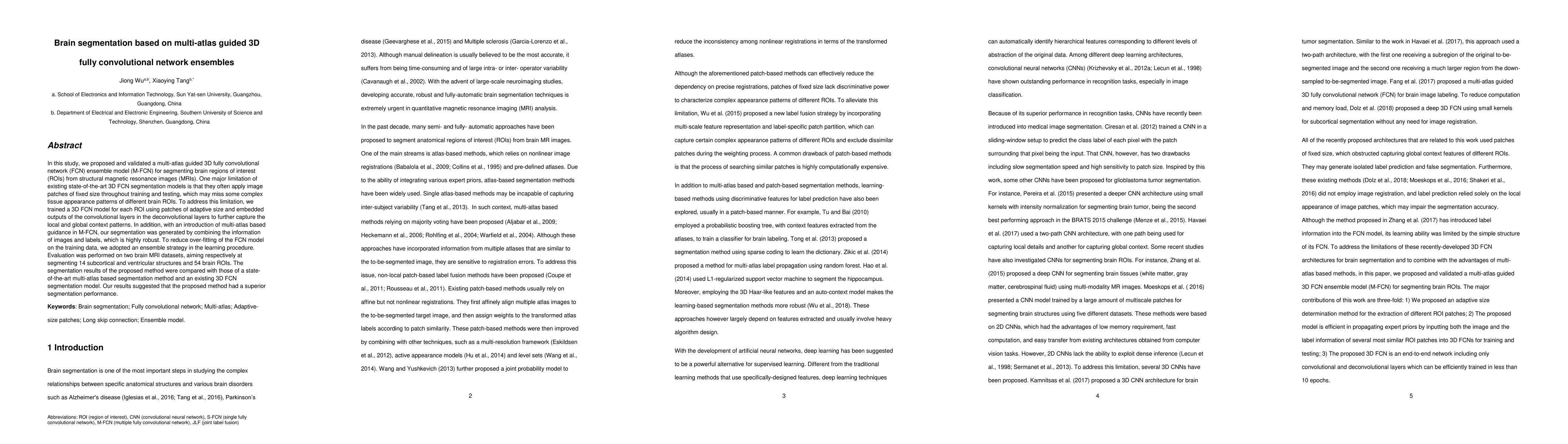
In this study, we proposed and validated a multi-atlas guided 3D fully convolutional network (FCN) ensemble model (M-FCN) for segmenting brain regions of interest (ROIs) from structural magnetic resonance images (MRIs). One major limitation of existing state-of-the-art 3D FCN segmentation models is that they often apply image patches of fixed size throughout training and testing, which may miss some complex tissue appearance patterns of different brain ROIs. To address this limitation, we trained a 3D FCN model for each ROI using patches of adaptive size and embedded outputs of the convolutional layers in the deconvolutional layers to further capture the local and global context patterns. In addition, with an introduction of multi-atlas based guidance in M-FCN, our segmentation was generated by combining the information of images and labels, which is highly robust. To reduce over-fitting of the FCN model on the training data, we adopted an ensemble strategy in the learning procedure. Evaluation was performed on two brain MRI datasets, aiming respectively at segmenting 14 subcortical and ventricular structures and 54 brain ROIs. The segmentation results of the proposed method were compared with those of a state-of-the-art multi-atlas based segmentation method and an existing 3D FCN segmentation model. Our results suggested that the proposed method had a superior segmentation performance.
Get AI-generated insights about this paper's methodology, results, and significance.
Current paper (gray), citations (green), references (blue)
Title | Authors | Year | Actions |
---|---|---|---|
Segmentation outperforms registration in quantitative analysis of brain iron
|
Junjie He, Bangkang Fu, Bo Wang, Zhenliang Xiong, Yunsong Peng, Rongpin Wang
|
2025
|
View Paper |
Masked Deformation Modeling for Volumetric Brain MRI Self-Supervised Pre-Training
|
Junyan Lyu, Perry F. Bartlett, Fatima A. Nasrallah, Xiaoying Tang
|
2024
|
View Paper |
Unfolding Explainable AI for Brain Tumor Segmentation
|
Muhammad Hassan, A. A. Fateh, Jieqiong Lin, Yijiang Zhuang, Guisen Lin, Hairui Xiong, You Zhou, Peiwu Qin, Hongwu Zeng
|
2024
|
View Paper |
Brain tumor segmentation in MRI with multi-modality spatial information enhancement and boundary shape correction
|
Zhiqin Zhu, Ziyu Wang, Guanqiu Qi, Neal Mazur, Pan Yang, Yu Liu
|
2024
|
View Paper |
Fast Diffeomorphic Image Registration using Patch based Fully Convolutional Networks
|
Jiong Wu, Shuang Zhou, Li Lin, Xin Wang, Wenxue Tan
|
2024
|
View Paper |
Reviewing 3D convolutional neural network approaches for medical image segmentation
|
A. Ilesanmi, Taiwo Ilesanmi, Babatunde O. Ajayi
|
2024
|
View Paper |
NnU-Net versus mesh growing algorithm as a tool for the robust and timely segmentation of neurosurgical 3D images in contrast-enhanced T1 MRI scans
|
Mathijs de Boer, Tessa M Kos, T. Fick, Jesse A M van Doormaal, Elisa Colombo, Hugo J. Kuijf, Pierre A. Robe, Luca Regli, L. Bartels, Tristan P C van Doormaal
|
2024
|
View Paper |
4T-Net: Multitask deep learning for nuclear analysis from pathology images
|
Vi Thi-Tuong Vo, Myung-Giun Noh, Soo-Hyung Kim
|
2024
|
View Paper |
Deep learning techniques for isointense infant brain tissue segmentation: a systematic literature review
|
S. T. Mhlanga, Serestina Viriri
|
2023
|
View Paper |
Toward hippocampal volume measures on ultra-high field magnetic resonance imaging: a comprehensive comparison study between deep learning and conventional approaches
|
Junyan Lyu, Perry F. Bartlett, Fatima A. Nasrallah, Xiaoying Tang
|
2023
|
View Paper |
MMAN-M2: Multiple multi-head attentions network based on encoder with missing modalities
|
Jiayao Li, Li Li, Ruizhi Sun, Gang Yuan, Shufan Wang, Shulin Sun
|
2023
|
View Paper |
Adaptive graph fusion learning for multi-view spectral clustering
|
Bo Zhou, Wenliang Liu, Meizhou Shen, Zhengyu Lu, Wenzhen Zhang, Luyun Zhang
|
2023
|
View Paper |
Improved segmentation of basal ganglia from MR images using convolutional neural network with crossover-typed skip connection
|
T. Sugino, Taichi Kin, Nobuhito Saito, Yoshikazu Nakajima
|
2023
|
View Paper |
Automatic semantic modeling of structured data sources with cross-modal retrieval
|
Rui-yun Xu, Wolfgang Mayer, Hailong Chu, Yitao Zhang, Hong-Yu Zhang, Yulong Wang, Youfa Liu, Zaiwen Feng
|
2023
|
View Paper |
Two-step multi-view data classification based on dynamic Graph-ELM
|
Li Li, Qihong Han, Jiayao Li, Zhanqi Cui
|
2023
|
View Paper |
Image-based MRI detection of brain tumours using convolutional neural networks
|
G. Preethi, NG Thangamma, S. P. Sankar, Md. Abul, Ala Walid, D. Suganthi, K. Deepthika
|
2023
|
View Paper |
EarlGAN: An enhanced actor-critic reinforcement learning agent-driven GAN for de novo drug design
|
Huidong Tang, Chen Li, Shuai Jiang, Huachong Yu, Sayaka Kamei, Yoshihiro Yamanishi, Yasuhiko Morimoto
|
2023
|
View Paper |
Multi-atlas subcortical segmentation: an orchestration of 3D fully convolutional network and generalized mixture function
|
Jiong Wu, Shuan He, Shuang Zhou
|
2023
|
View Paper |
Thalamus Segmentation Using Deep Learning with Diffusion MRI Data: An Open Benchmark
|
G. Pinheiro, Lorenza Brusini, D. Carmo, Renata Prôa, Thays Abreu, Simone Appenzeller, G. Menegaz, Letícia Rittner
|
2023
|
View Paper |
Arbitrary Order Total Variation for Deformable Image Registration
|
Jinming Duan, Xi Jia, Joseph Bartlett, Wenqi Lu, Zhaowen Qiu
|
2023
|
View Paper |
Accurate segmentation of neonatal brain MRI with deep learning
|
Leonie Richter, Ahmed E. Fetit
|
2022
|
View Paper |
Application of medical imaging methods and artificial intelligence in tissue engineering and organ-on-a-chip
|
Wanying Gao, Chunyan Wang, Qiwei Li, Xijing Zhang, Jianmin Yuan, Dianfu Li, Yu Sun, Zaozao Chen, Zhongze Gu
|
2022
|
View Paper |
DS3-Net: Difficulty-perceived Common-to-T1ce Semi-Supervised Multimodal MRI Synthesis Network
|
Ziqi Huang, Li Lin, Pujin Cheng, Kai Pan, Xiaoying Tang
|
2022
|
View Paper |
Methods for segmenting cracks in 3d images of concrete: A comparison based on semi-synthetic images
|
Tin Barisin, Christian Jung, Franziska Müsebeck, C. Redenbach, K. Schladitz
|
2021
|
View Paper |
Brain tumor segmentation based on the dual-path network of multi-modal MRI images
|
Lingling Fang, Xin Wang
|
2021
|
View Paper |
Brain Tumor Segmentation From Multi-Modal MR Images via Ensembling UNets
|
Yue Zhang, Pinyuan Zhong, Dabin Jie, Jiewei Wu, Shan Zeng, Jianping Chu, Yilong Liu, E. Wu, Xiaoying Tang
|
2021
|
View Paper |
Characterization of the volume and thickness of DIEP flap by CTA image processing
|
María Alejandra Cerón, Julián D. Loaiza Duque, S. B. Verdoy, Ana López Ojeda, Marta Alcover Morro, Pedro Quetglas Barea, H. Rojas, A. J. S. Egea
|
2021
|
View Paper |
System for Predicting the Origin of Neurological Symptomatics in Children after an Ischemic Stroke
|
G. Mashevskiy, Polina Dubrovina
|
2020
|
View Paper |
AssemblyNet: A large ensemble of CNNs for 3D whole brain MRI segmentation
|
P. Coupé, Boris Mansencal, Michaël Clément, Rémi Giraud, B. D. D. Senneville, T. Thong, V. Lepetit, J. Manjón
|
2019
|
View Paper |
Consistent Segmentation of Longitudinal Brain MR Images with Spatio-Temporal Constrained Networks
|
Jie Wei, F. Shi, Zhiming Cui, Yongsheng Pan, Yong Xia, D. Shen
|
2021
|
View Paper |
Comments (0)