BrainPainter is a versatile software for visualizing brain structures and pathological processes, offering compatibility with any neuroimaging analysis tools and the ability to generate dynamic visualizations. It leverages Blender's powerful 3D graphics engine for advanced visualizations, and is accessible via web browser or Docker container.
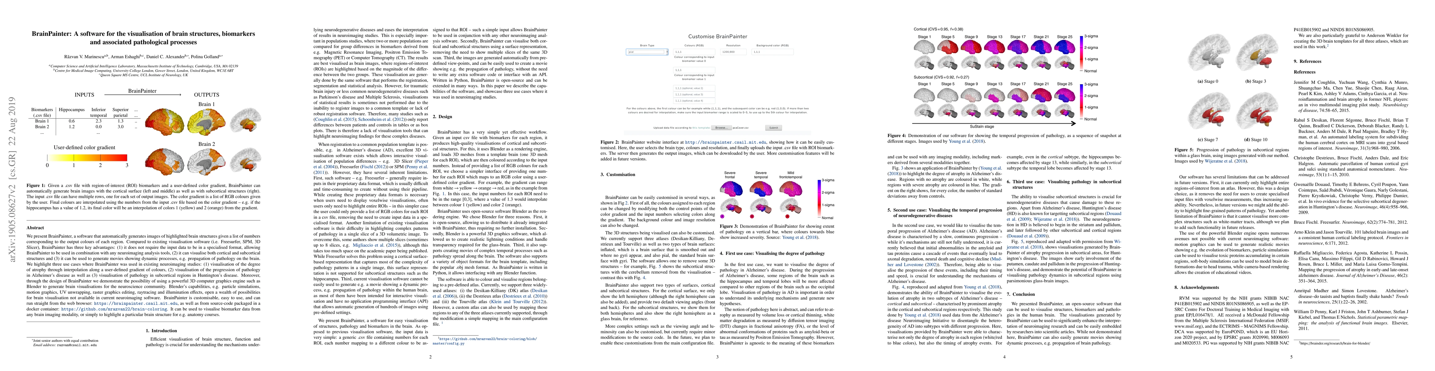
We present BrainPainter, a software that automatically generates images of highlighted brain structures given a list of numbers corresponding to the output colours of each region. Compared to existing visualisation software (i.e. Freesurfer, SPM, 3D Slicer), BrainPainter has three key advantages: (1) it does not require the input data to be in a specialised format, allowing BrainPainter to be used in combination with any neuroimaging analysis tools, (2) it can visualise both cortical and subcortical structures and (3) it can be used to generate movies showing dynamic processes, e.g. propagation of pathology on the brain. We highlight three use cases where BrainPainter was used in existing neuroimaging studies: (1) visualisation of the degree of atrophy through interpolation along a user-defined gradient of colours, (2) visualisation of the progression of pathology in Alzheimer's disease as well as (3) visualisation of pathology in subcortical regions in Huntington's disease. Moreover, through the design of BrainPainter we demonstrate the possibility of using a powerful 3D computer graphics engine such as Blender to generate brain visualisations for the neuroscience community. Blender's capabilities, e.g. particle simulations, motion graphics, UV unwrapping, raster graphics editing, raytracing and illumination effects, open a wealth of possibilities for brain visualisation not available in current neuroimaging software. BrainPainter is customisable, easy to use, and can run straight from the web browser: https://brainpainter.csail.mit.edu , as well as from source-code packaged in a docker container: https://github.com/mrazvan22/brain-coloring . It can be used to visualise biomarker data from any brain imaging modality, or simply to highlight a particular brain structure for e.g. anatomy courses.
This graph shows how this paper is connected to other research. The current paper is shown in dark gray, papers that cite it are in green, and papers it references are in blue. You can:
• Hover over any node to see the paper title
• Click a node to view that paper's details
• Drag nodes to rearrange the graph
• Use the zoom controls (bottom right) to zoom in/out
Title | Authors | Year | Actions |
---|---|---|---|
Neuroimaging-based data-driven subtypes of spatiotemporal atrophy due to Parkinson’s disease
|
Zeena Shawa, C. Shand, Beatrice Taylor, H. Berendse, C. Vriend, T. D. van Balkom, O. A. van den Heuvel, Y. D. van der Werf, Jiun-jie Wang, Chih-Chien Tsai, Jason Druzgal, Benjamin Newman, Tracy R. Melzer, T. Pitcher, J. Dalrymple-Alford, Tim J Anderson, Gaëtan Garraux, Mario Rango, P. Schwingenschuh, Melanie Suette, Laura M. Parkes, Sarah Al-Bachari, Johannes Klein, Michele T M Hu, Corey T McMillan, F. Piras, D. Vecchio, Clelia Pellicano, Chengcheng Zhang, K. Poston, Elnaz Ghasemi, F. Cendes, Clarissa Yasuda, D. Tosun, Philip Mosley, Paul M. Thompson, N. Jahanshad, Conor Owens-Walton, Emile d’Angremont, Eva M. van Heese, Max A. Laansma, André Altmann, Max A. Laansma, Joanna K Bright, Sarah Al-Bachari, Tim J Anderson, Tyler Ard, F. Assogna, K. Baquero, H. Berendse, Benajmin Newman, F. Cendes, J. Dalrymple-Alford, R. D. de Bie, I. Debove, M. Dirkx, Jason Druzgal, H. Emsley, Gaëtan Garraux, R. Guimaraes, B. Gutman, R. C. Helmich, Johannes Klein, Clare E Mackay, Corey T McMillan, Tracy R. Melzer, Laura M. Parkes, F. Piras, T. Pitcher, K. Poston, Mario Rango, L. Ribeiro, Cristiane S Rocha, Christian Rummel, Lucas S R Santos, Reinhold Schmidt, P. Schwingenschuh, Gianfranco Spalletta, L. Squarcina, O. A. van den Heuvel, C. Vriend, Jiun-jie Wang, D. Weintraub, R. Wiest, Clarissa Yasuda, N. Jahanshad, Paul M. Thompson, Y. D. van der Werf, Rimona S Weil, Neil P. Oxtoby
|
2025
|
View Paper |
Putative Mechanisms of Electroconvulsive Therapy in Treatment Resistant Schizophrenia Examined using Magnetic Resonance Imaging
|
Neelabja Roy, D. Ithal, U. Mehta, R. Basavaraju, R. D. Bharath, Nicolas R Bolo, J. Thirthalli, B. Gangadhar, M. Keshavan
|
2025
|
View Paper |
OCL: Ordinal Contrastive Learning for Imputating Features with Progressive Labels
|
Seunghun Baek, Jaeyoon Sim, Guorong Wu, Won Hwa Kim
|
2025
|
View Paper |
Cortical thinning in temporal pole, a core region in Alzheimer’s disease, in non-demented, middle-aged APOE-ε4 and PICALM-AA/AG carriers
|
P. Dzianok, Jakub Wojciechowski, Tomasz Wolak, E. Kublik
|
2025
|
View Paper |
Distinct spatiotemporal atrophy patterns in corticobasal syndrome are associated with different underlying pathologies
|
W. J. Scotton, C. Shand, E. Todd, M. Bocchetta, C. Kobylecki, D. M. Cash, L. Vandevrede, H. Heuer, Annelies Quaegebeur, A. Young, Neil Oxtoby, Daniel C Alexander, James B. Rowe, H. Morris, Adam L Boxer, J. Rohrer, P. Wijeratne
|
2025
|
View Paper |
A data-driven framework for biomarker discovery applied to optimizing modern clinical and preclinical trials on Alzheimer’s disease
|
Isaac Llorente-Saguer, Neil P. Oxtoby
|
2024
|
View Paper |
Beyond the Buzz: Cortical and subcortical brain changes in patients with pulsatile tinnitus
|
J. Remer, K. Narsinh, Travis Caton, A. Lamboy, A. Tu-Chan, Ashish Raj, M. Amans
|
2024
|
View Paper |
Autonomic biosignals, seizure detection, and forecasting.
|
Gadi Miron, M. Halimeh, J. Jeppesen, Tobias Loddenkemper, C. Meisel
|
2024
|
View Paper |
Modality-Agnostic Style Transfer for Holistic Feature Imputation
|
Seunghun Baek, Jaeyoon Sim, Mustafa Dere, Minjeong Kim, Guorong Wu, Won Hwa Kim
|
2024
|
View Paper |
Data-driven neuroanatomical subtypes of primary progressive aphasia
|
Beatrice Taylor, M. Bocchetta, Cameron Shand, E. Todd, Anthipa Chokesuwattanaskul, S. Crutch, Jason D. Warren, J. Rohrer, Chris J D Hardy, Neil P. Oxtoby
|
2024
|
View Paper |
Distinct spatiotemporal atrophy patterns in corticobasal syndrome are associated with different underlying pathologies
|
W. J. Scotton, C. Shand, E. Todd, M. Bocchetta, D. M. Cash, L. Vandevrede, H. Heuer, A. Young, N. Oxtoby, D. Alexander, J. Rowe, H. Morris, A. Boxer, J. D. Rohrer, P. Wijeratne
|
2024
|
View Paper |
Brain morphological variability between whites and African Americans: the importance of racial identity in brain imaging research
|
Daniel Atilano‐Barbosa, Fernando A. Barrios
|
2023
|
View Paper |
Brain MRI changes in degenerative cervical myelopathy: a systematic review
|
Amir Rafati Fard, O. Mowforth, M. Yuan, Samuel Myrtle, K. Lee, Arka Banerjee, Maaz Khan, M. Kotter, V. Newcombe, E. Stamatakis, B. Davies
|
2023
|
View Paper |
Deep Geometric Learning With Monotonicity Constraints for Alzheimer’s Disease Progression
|
Seungwoo Jeong, Wonsik Jung, Junghyo Sohn, Heung-Il Suk
|
2023
|
View Paper |
Quantifying the Growth of Glioblastoma Tumors Using Multimodal MRI Brain Images
|
Anisha Das, Shengxian Ding, Rongjie Liu, Chao Huang
|
2023
|
View Paper |
Cortical thickness moderates intraindividual variability in prefrontal cortex activation patterns of older adults during walking
|
Daliah Ross, M. Wagshul, M. Izzetoglu, R. Holtzer
|
2023
|
View Paper |
Learning Covariance-Based Multi-Scale Representation of Neuroimaging Measures for Alzheimer Classification
|
Seung-Bin Baek, Injun Choi, Mustafa Dere, Minjeong Kim, Guorong Wu, Won Hwa Kim
|
2023
|
View Paper |
Default mode network failure and neurodegeneration across aging and amnestic and dysexecutive Alzheimer’s disease
|
Nick Corriveau-Lecavalier, J. Gunter, Michael Kamykowski, E. Dicks, H. Botha, W. Kremers, J. Graff‐Radford, D. Wiepert, C. Schwarz, E. Yacoub, D. Knopman, B. Boeve, K. Uğurbil, R. Petersen, C. Jack, Melissa Terpstra, David T. Jones
|
2023
|
View Paper |
Crop filling: A pipeline for repairing memory clinic MRI corrupted by partial brain coverage
|
G. Leal, T. Whitfield, Janaki Praharaju, Zuzana Walker, N. Oxtoby
|
2023
|
View Paper |
Neural Correlates of Delay Discounting in the Light of Brain Imaging and Non-Invasive Brain Stimulation: What We Know and What Is Missed
|
A. Moro, Daniele Saccenti, Mattia Ferro, S. Scaini, A. Malgaroli, J. Lamanna
|
2023
|
View Paper |
Telomere length is causally connected to brain MRI image derived phenotypes: A mendelian randomization study
|
Ahmed M. A. Salih, I. Galazzo, Steffen E. Petersen, K. Lekadir, P. Radeva, G. Menegaz, A. Altmann
|
2022
|
View Paper |
Relationship of serum beta‐synuclein with blood biomarkers and brain atrophy
|
P. Oeckl, S. Anderl-Straub, A. Danek, J. Diehl-Schmid, K. Fassbender, K. Fliessbach, S. Halbgebauer, H. Huppertz, H. Jahn, J. Kassubek, J. Kornhuber, B. Landwehrmeyer, M. Lauer, J. Prudlo, A. Schneider, M. Schroeter, P. Steinacker, A. Volk, M. Wagner, J. Winkelmann, J. Wiltfang, A. Ludolph, M. Otto
|
2022
|
View Paper |
Longitudinal study of early mild cognitive impairment via similarity-constrained group learning and self-attention based SBi-LSTM
|
Baiying Lei, Yuwen Zhang, Dongdong Liu, Yanwu Xu, Guanghui Yue, Jiuwen Cao, Huoyou Hu, Shuangzhi Yu, Peng Yang, Tianfu Wang, Yali Qiu, Xiaohua Xiao, Shuqiang Wang
|
2022
|
View Paper |
Disentangled Representation of Longitudinal Β-Amyloid for AD Via Sequential Graph Variational Autoencoder with Supervision
|
Fan Yang, Guorong Wu, Won Hwa Kim
|
2022
|
View Paper |
Regional cerebral hypometabolism on 18F‐FDG PET/CT scan in delirium is independent of acute illness and dementia
|
Anita Nitchingham, J. V. Pereira, E. Wegner, Vincent Oxenham, Jacqueline F. Close, G. Caplan
|
2022
|
View Paper |
Prefrontal cortex activation during dual-task walking in older adults is moderated by thickness of several cortical regions
|
Daliah Ross, M. Wagshul, M. Izzetoglu, R. Holtzer
|
2021
|
View Paper |
Characterizing the Clinical Features and Atrophy Patterns of MAPT-Related Frontotemporal Dementia With Disease Progression Modeling
|
A. Young, M. Bocchetta, L. Russell, R. Convery, Georgia Peakman, E. Todd, D. Cash, C. Greaves, J. V. van Swieten, L. Jiskoot, H. Seelaar, F. Moreno, Raquel Sánchez-Valle, B. Borroni, R. Laforce, M. Masellis, M. Tartaglia, C. Graff, D. Galimberti, J. Rowe, E. Finger, M. Synofzik, R. Vandenberghe, A. de Mendonça, F. Tagliavini, I. Santana, S. Ducharme, C. Butler, A. Gerhard, J. Levin, A. Danek, M. Otto, S. Sorbi, S. C. Williams, D. Alexander, J. Rohrer
|
2021
|
View Paper |
Progression along data-driven disease timelines is predictive of Alzheimer’s disease in a population-based cohort
|
Vikram Venkatraghavan, E. Vinke, E. Bron, W. Niessen, M. Ikram, S. Klein, M. Vernooij
|
2021
|
View Paper |
Regional grey matter microstructural changes and volume loss according to disease duration in multiple sclerosis patients
|
E. Solana, E. Martínez-Heras, Víctor Montal, Eduard Vilaplana, E. Lopez-Soley, J. Raduà, N. Solà-Valls, C. Montejo, Y. Blanco, I. Pulido-Valdeolivas, M. Sepúlveda, Magí Andorrà, J. Berenguer, P. Villoslada, Martínez-Lapiscina Eh, F. Prados, A. Saiz, J. Fortea, S. Llufriu
|
2021
|
View Paper |
Investigating hypotheses of neurodegeneration by learning dynamical systems of protein propagation in the brain
|
Sara Garbarino, Marco Lorenzi, Alzheimer's Disease Neuroimaging Initiative
|
2021
|
View Paper |
Simulating the outcome of amyloid treatments in Alzheimer's disease from imaging and clinical data
|
Clément Abi Nader, N. Ayache, G. Frisoni, P. Robert, Marco Lorenzi
|
2021
|
View Paper |
Increased facial asymmetry in focal epilepsies associated with unilateral lesions
|
S. Balestrini, Seymour M Lopez, K. Chinthapalli, Narek Sargsyan, R. Demurtas, S. Vos, A. Altmann, M. Suttie, P. Hammond, S. Sisodiya
|
2021
|
View Paper |
BrainPainter v2: Mouse Brain Visualization Software
|
Vedvayas Mallela, P. Golland, Razvan V. Marinescu
|
2021
|
View Paper |
Modeling seeding and neuroanatomic spread of pathology in amyotrophic lateral sclerosis
|
S. Pandya, P. Maia, Benjamin Freeze, R. Menke, K. Talbot, M. Turner, A. Raj
|
2021
|
View Paper |
Resilience, pain, and the brain: Relationships differ by sociodemographics
|
Jared J. Tanner, Alisa J. Johnson, Ellen L. Terry, J. Cardoso, Cynthia Garvan, R. Staud, G. Deutsch, H. Deshpande, S. Lai, A. Addison, D. Redden, B. Goodin, C. Price, R. Fillingim, K. Sibille
|
2021
|
View Paper |
Tau-first subtype of Alzheimer’s disease consistently identified across in vivo and post mortem studies
|
L. Aksman, N. Oxtoby, M. A. Scelsi, P. Wijeratne, A. Young, I. L. Alves, F. Barkhof, D. Alexander, A. Altmann
|
2020
|
View Paper |
Simulating the outcome of amyloid treatments in Alzheimer’s Disease from multi-modal imaging and clinical data
|
Clément Abi Nader, N. Ayache, G. Frisoni, P. Robert, Marco Lorenzi
|
2020
|
View Paper |
Cross-Disorder Analysis of Brain Structural Abnormalities in Six Major Psychiatric Disorders: A Secondary Analysis of Mega- and Meta-analytical Findings From the ENIGMA Consortium
|
N. Opel, J. Goltermann, M. Hermesdorf, K. Berger, B. Baune, U. Dannlowski
|
2020
|
View Paper |
Deep recurrent model for individualized prediction of Alzheimer’s disease progression
|
Wonsik Jung, E. Jun, Heung-Il Suk, Alzheimer's Disease Neuroimaging Initiative
|
2020
|
View Paper |
Deep Recurrent Disease Progression Model for Conversion-Time Prediction of Alzheimer's Disease
|
Wonsik Jung, E. Jun, Heung-Il Suk
|
2020
|
View Paper |
ADM IRE ++: Explainable Anomaly Detection in the Human Brain via Inductive Learning on Temporal Multiplex Networks
|
Ali Behrouz, Margo Seltzer
|
2023
|
View Paper |
Anomaly Detection in Human Brain via Inductive Learning on Temporal Multiplex Networks
|
Ali Behrouz, Margo Seltzer
|
2023
|
View Paper |
OP-BRCM210068 1..14
|
S. Balestrini, Seymour M Lopez, K. Chinthapalli, Narek Sargsyan, R. Demurtas, S. Vos, A. Altmann, M. Suttie, P. Hammond, S. Sisodiya
|
2021
|
View Paper |
Published: 2023-09-15
Thomas Wahl, Michel Duprez, Joséphine Riedinger, Axel Hutt
Published: 2023-03-21
José Gerardo Suárez-García, Benito de Celis-Alonso, María Isabel Antonio-de la Rosa, Nora Coral Soriano-Becerril, Javier M. Hernández-López
Published: 2024-09-07
Comments (0)