AI Quick Summary
A new brain lesion segmentation model called BrainSegDMLF overcomes limitations of existing methods by integrating multi-modal data, detecting small lesions with limited data, and automatically generating segmentation masks.
Quick Answers
What is "BrainSegDMlF: A Dynamic Fusion-enhanced SAM for Brain Lesion Segmentation" about?
A new brain lesion segmentation model called BrainSegDMLF overcomes limitations of existing methods by integrating multi-modal data, detecting small lesions with limited data, and automatically generating segmentation masks.
What methodology did the authors use?
The research introduces BrainSegDMLF, a large-scale automated segmentation model for brain lesions, featuring a Dynamic Modal Interactive Fusion (DMIF) module for multi-modal data integration, a Layer-by-Layer Upsampling Decoder for feature extraction, and automatic segmentation masks. More in Methodology →
What are the key results?
BrainSegDMLF effectively integrates multi-modal data for comprehensive lesion information acquisition. — The model can detect small lesions even with limited data through rich feature extraction. More in Key Results →
Why is this work significant?
This research is significant as it addresses the challenges of existing brain lesion segmentation methods by providing a fully automated model that utilizes multi-modal data and generates accurate segmentation masks, enhancing diagnostic efficiency. More in Significance →
What are the main limitations?
The paper does not explicitly mention specific limitations of the proposed method. — Performance under diverse pathological conditions and with various imaging modalities might not be addressed. More in Limitations →
Abstract
The segmentation of substantial brain lesions is a significant and challenging task in the field of medical image segmentation. Substantial brain 
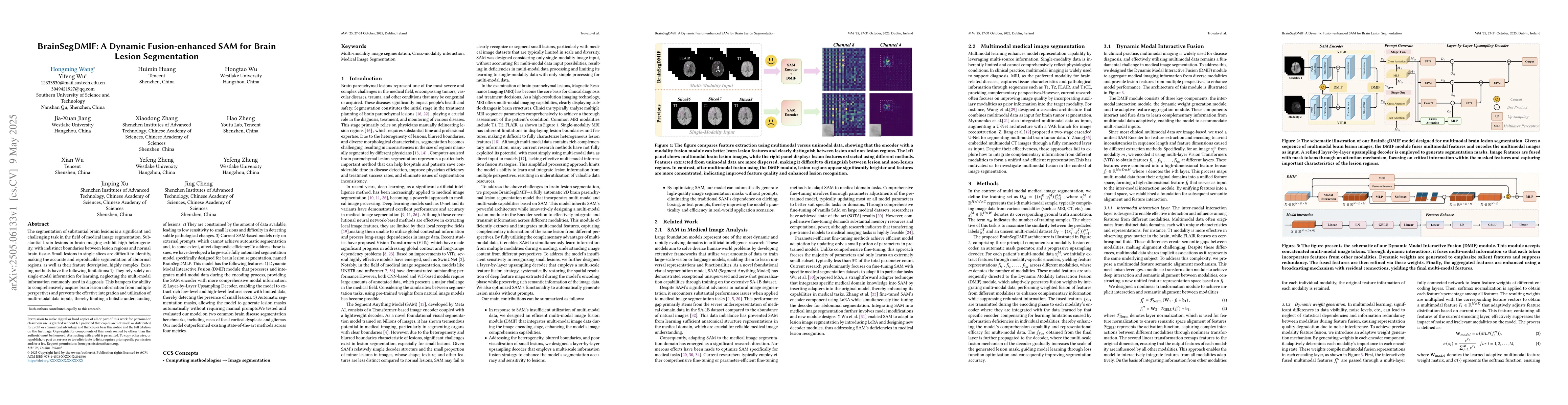
lesions in brain imaging exhibit high heterogeneity, with indistinct boundaries between lesion regions and normal brain tissue. Small lesions in single slices are difficult to identify, making the accurate and reproducible segmentation of abnormal regions, as well as their feature description, highly complex. Existing methods have the following limitations: 1) They rely solely on single-modal information for learning, neglecting the multi-modal information commonly used in diagnosis. This hampers the ability to comprehensively acquire brain lesion information from multiple perspectives and prevents the effective integration and utilization of multi-modal data inputs, thereby limiting a holistic understanding of lesions. 2) They are constrained by the amount of data available, leading to low sensitivity to small lesions and difficulty in detecting subtle pathological changes. 3) Current SAM-based models rely on external prompts, which cannot achieve automatic segmentation and, to some extent, affect diagnostic efficiency.To address these issues, we have developed a large-scale fully automated segmentation model specifically designed for brain lesion segmentation, named BrainSegDMLF. This model has the following features: 1) Dynamic Modal Interactive Fusion (DMIF) module that processes and integrates multi-modal data during the encoding process, providing the SAM encoder with more comprehensive modal information. 2) Layer-by-Layer Upsampling Decoder, enabling the model to extract rich low-level and high-level features even with limited data, thereby detecting the presence of small lesions. 3) Automatic segmentation masks, allowing the model to generate lesion masks automatically without requiring manual prompts.
AI Key Findings
Generated Jun 08, 2025
Methodology — What approach did the authors take?
The research introduces BrainSegDMLF, a large-scale automated segmentation model for brain lesions, featuring a Dynamic Modal Interactive Fusion (DMIF) module for multi-modal data integration, a Layer-by-Layer Upsampling Decoder for feature extraction, and automatic segmentation masks.
Key Results — What are the main findings?
- BrainSegDMLF effectively integrates multi-modal data for comprehensive lesion information acquisition.
- The model can detect small lesions even with limited data through rich feature extraction.
Significance — Why does this research matter?
This research is significant as it addresses the challenges of existing brain lesion segmentation methods by providing a fully automated model that utilizes multi-modal data and generates accurate segmentation masks, enhancing diagnostic efficiency.
Technical Contribution — What is the technical contribution?
The main technical contribution is the Dynamic Modal Interactive Fusion (DMIF) module and the Layer-by-Layer Upsampling Decoder, which enable effective multi-modal data integration and feature extraction for brain lesion segmentation.
Novelty — What is new about this work?
BrainSegDMLF stands out by introducing an automated, multi-modal fusion approach for brain lesion segmentation, addressing the limitations of existing single-modal and prompt-dependent models.
Limitations — What are the limitations of this study?
- The paper does not explicitly mention specific limitations of the proposed method.
- Performance under diverse pathological conditions and with various imaging modalities might not be addressed.
Future Work — What did the authors propose for future work?
- Explore the model's performance with additional imaging modalities and pathological variations.
- Investigate the model's applicability to other types of medical image segmentation tasks.
How to Cite This Paper
@article{zheng2025brainsegdmlf,
title = {BrainSegDMlF: A Dynamic Fusion-enhanced SAM for Brain Lesion
Segmentation},
author = {Zheng, Hao and Wang, Hongming and Wu, Hongtao and others},
year = {2025},
eprint = {2505.06133},
archivePrefix = {arXiv},
primaryClass = {cs.CV},
}
Zheng, H., Wang, H., Wu, H., Huang, H., Jiang, J., Cheng, J., Xu, J., Wu, X., Zhang, X., Zheng, Y., & Wu, Y. (2025). BrainSegDMlF: A Dynamic Fusion-enhanced SAM for Brain Lesion
Segmentation. arXiv. https://arxiv.org/abs/2505.06133
Zheng, Hao, et al. "BrainSegDMlF: A Dynamic Fusion-enhanced SAM for Brain Lesion
Segmentation." arXiv, 2025, arxiv.org/abs/2505.06133.
PDF Preview
Citation Network
Current paper (gray), citations (green), references (blue)
Display is limited for performance on very large graphs.
Similar Papers
Found 4 papersMorphology-Enhanced CAM-Guided SAM for weakly supervised Breast Lesion Segmentation
Xin Yue, Xiaoling Liu, Qing Zhao et al.
Unsupervised Domain Adaptation with SAM-RefiSeR for Enhanced Brain Tumor Segmentation
Dillan Imans, Phuoc-Nguyen Bui, Duc-Tai Le et al.
Ultrasound SAM Adapter: Adapting SAM for Breast Lesion Segmentation in Ultrasound Images
Zhengzheng Tu, Le Gu, Xixi Wang et al.
LCAUnet: A skin lesion segmentation network with enhanced edge and body fusion
Qisen Ma, Keming Mao, Gao Wang et al.
No citations found for this paper.
Comments (0)