AI Quick Summary
This review examines recent advancements in fluorescence microscopy and tissue engineering techniques to enhance high-resolution sub-cellular imaging in thick 3D engineered tissues, offering researchers a comprehensive overview of available imaging options and recent breakthroughs in overcoming traditional imaging limitations.
Quick Answers
What is "Bridging high resolution sub-cellular imaging with physiologically relevant engineered tissues" about?
This review examines recent advancements in fluorescence microscopy and tissue engineering techniques to enhance high-resolution sub-cellular imaging in thick 3D engineered tissues, offering researchers a comprehensive overview of available imaging options and recent breakthroughs in overcoming traditional imaging limitations.
What methodology did the authors use?
This research reviews advancements in fluorescence microscopy, various fluorescent probes, and material processing techniques to overcome challenges in obtaining high-resolution images from thick 3D engineered tissues. More in Methodology →
What are the key results?
Multiphoton microscopy allows for high-resolution imaging of thick, scattering biological specimens with minimal distortion. — Light-sheet fluorescence microscopy provides high-resolution, three-dimensional views of biological samples, especially thick 3D engineered tissues, with minimal photodamage. More in Key Results →
Why is this work significant?
This research is crucial for tissue engineering as it enables detailed visualization of subcellular structures within 3D tissues, providing insights into critical cellular processes and their responses to engineered environments. More in Significance →
What are the main limitations?
Multiphoton microscopy can lead to increased photobleaching and photodamage in the focal plane, especially with high-intensity near-infrared pulses. — Light-sheet microscopy requires samples to be transparent enough for the light-sheet to penetrate, often necessitating time-consuming clearing techniques that may alter the sample's natural state. More in Limitations →
Abstract
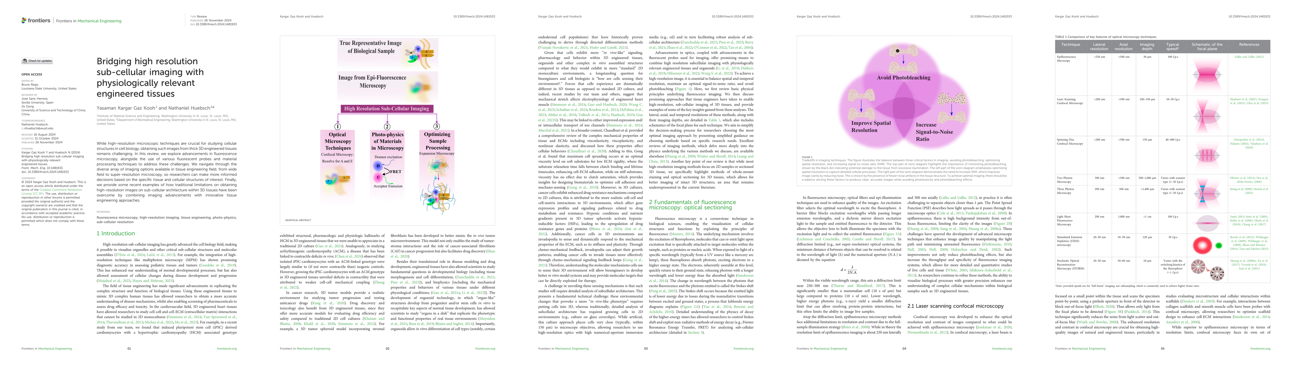
While high-resolution microscopic techniques are crucial for studying cellular structures in cell biology, obtaining such images from thick 3D engineered tissues remains challenging. In this review, we explore advancements in fluorescence microscopy, alongside the use of various fluorescent probes and material processing techniques to address these challenges. We navigate through the diverse array of imaging options available in tissue engineering field, from wide field to super-resolution microscopy, so researchers can make more informed decisions based on the specific tissue and cellular structures of interest. Finally, we provide some recent examples of how traditional limitations on obtaining high-resolution images on sub-cellular architecture within 3D tissues have been overcome by combining imaging advancements with innovative tissue engineering approaches.
AI Key Findings
Generated Jun 11, 2025
Methodology — What approach did the authors take?
This research reviews advancements in fluorescence microscopy, various fluorescent probes, and material processing techniques to overcome challenges in obtaining high-resolution images from thick 3D engineered tissues.
Key Results — What are the main findings?
- Multiphoton microscopy allows for high-resolution imaging of thick, scattering biological specimens with minimal distortion.
- Light-sheet fluorescence microscopy provides high-resolution, three-dimensional views of biological samples, especially thick 3D engineered tissues, with minimal photodamage.
- STED microscopy surpasses traditional diffraction limits, enabling super-resolution imaging beyond the 200-300 nm limit of conventional light microscopes.
- FRET, FLIM, and FRAP microscopy techniques leverage photophysical properties of fluorescence molecules to surpass the diffraction limit and study molecular dynamics within live cells.
- STORM provides high spatial resolution and does not require high-intensity or continuous illumination of lasers, minimizing photodamage to samples.
Significance — Why does this research matter?
This research is crucial for tissue engineering as it enables detailed visualization of subcellular structures within 3D tissues, providing insights into critical cellular processes and their responses to engineered environments.
Technical Contribution — What is the technical contribution?
The review presents a comprehensive analysis of various microscopy techniques, fluorescent probes, and material processing methods to enhance subcellular imaging in thick 3D engineered tissues.
Novelty — What is new about this work?
This research distinguishes itself by integrating advancements in fluorescence microscopy, fluorescent probes, and material processing techniques, providing a detailed comparison and guidance for researchers in the tissue engineering field.
Limitations — What are the limitations of this study?
- Multiphoton microscopy can lead to increased photobleaching and photodamage in the focal plane, especially with high-intensity near-infrared pulses.
- Light-sheet microscopy requires samples to be transparent enough for the light-sheet to penetrate, often necessitating time-consuming clearing techniques that may alter the sample's natural state.
- STED microscopy can cause increased photodamage, photobleaching, and phototoxic effects due to prolonged exposure of biological samples to intense light.
Future Work — What did the authors propose for future work?
- Further optimization of excitation wavelengths, photon numbers, and laser sources in multiphoton microscopy to maximize penetration depth and minimize photodamage.
- Development of more efficient and less invasive sample clearing techniques for light-sheet microscopy.
- Improvement of STED microscopy to reduce photodamage and increase temporal resolution.
How to Cite This Paper
@article{huebsch2025bridging,
title = {Bridging high resolution sub-cellular imaging with physiologically
relevant engineered tissues},
author = {Huebsch, Nathaniel and Kooh, Yasaman Kargar Gaz},
year = {2025},
eprint = {2502.03661},
archivePrefix = {arXiv},
primaryClass = {q-bio.TO},
doi = {10.3389/fmech.2024.1481933},
}
Huebsch, N., & Kooh, Y. (2025). Bridging high resolution sub-cellular imaging with physiologically
relevant engineered tissues. arXiv. https://doi.org/10.3389/fmech.2024.1481933
Huebsch, Nathaniel, and Yasaman Kargar Gaz Kooh. "Bridging high resolution sub-cellular imaging with physiologically
relevant engineered tissues." arXiv, 2025, doi.org/10.3389/fmech.2024.1481933.
PDF Preview
Citation Network
Current paper (gray), citations (green), references (blue)
Display is limited for performance on very large graphs.
Similar Papers
Found 4 papersHigh SNR 3D Imaging from Millimeter-scale Thick Tissues to Cellular Dynamics via Structured Illumination Microscopy
Mengrui Wang, Manming Shu, Jiajing Yan et al.
No citations found for this paper.
Comments (0)