Imaging protein interactions in vivo with sub-cellular resolution
Publication
Metrics
AI Quick Summary
This paper presents a method for imaging protein interactions in living cells using a spectrally-resolved two-photon microscope, enabling sub-cellular resolution. This approach aims to track the dynamics of protein complexes without the need for sequential excitation, potentially offering more accurate and real-time insights into cellular processes.
Paper Preview
Abstract
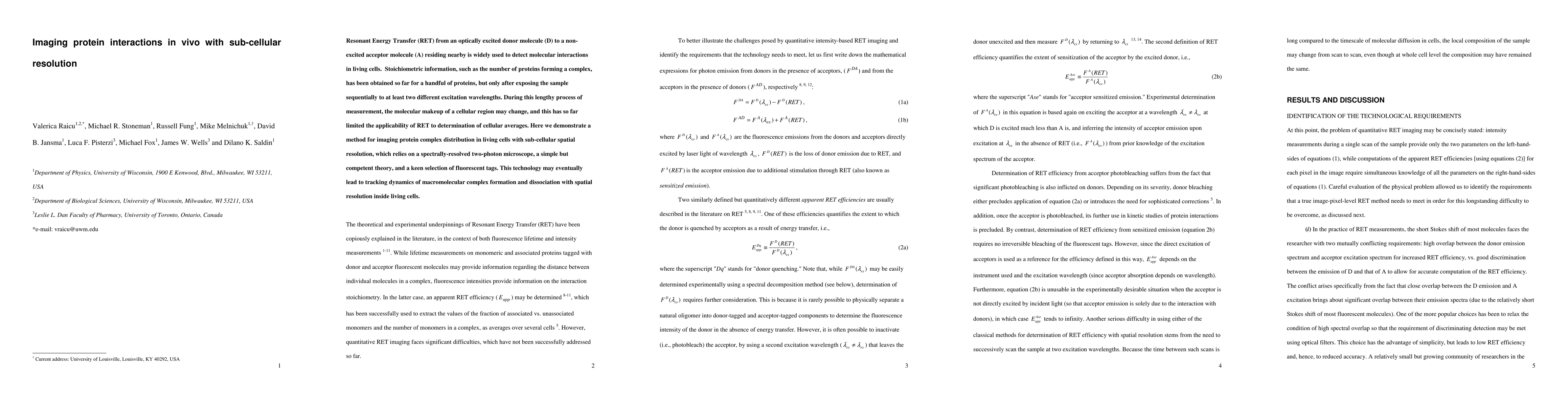
Resonant Energy Transfer (RET) from an optically excited donor molecule (D) to a non-excited acceptor molecule (A) residing nearby is widely used to detect molecular interactions in living cells. Stoichiometric information, such as the number of proteins forming a complex, has been obtained so far for a handful of proteins, but only after exposing the sample sequentially to at least two different excitation wavelengths. During this lengthy process of measurement, the molecular makeup of a cellular region may change, and this has so far limited the applicability of RET to determination of cellular averages. Here we demonstrate a method for imaging protein complex distribution in living cells with sub-cellular spatial resolution, which relies on a spectrally-resolved two-photon microscope, a simple but competent theory, and a keen selection of fluorescent tags. This technology may eventually lead to tracking dynamics of macromolecular complex formation and dissociation with spatial resolution inside living cells.
AI Key Findings
Get AI-generated insights about this paper's methodology, results, significance, and more — seven facets brought into focus.
Impact
Paper Details
PDF Preview
Key Terms
Citation Network
Current paper (gray), citations (green), references (blue)
Display is limited for performance on very large graphs.
Discussion 0