Bridging the Gap Between 2D and 3D Organ Segmentation with Volumetric Fusion Net

Source: ArXiv

AI Quick Summary

This paper proposes a framework that uses 2D networks trained on different viewpoints to segment organs and a 3D Volumetric Fusion Net (VFN) to combine these results, achieving higher efficiency and accuracy compared to traditional 2D and 3D segmentation methods. The authors introduce a cross-cross-augmentation strategy to optimize training with limited data.

Share: Share on X LinkedIn

Summary

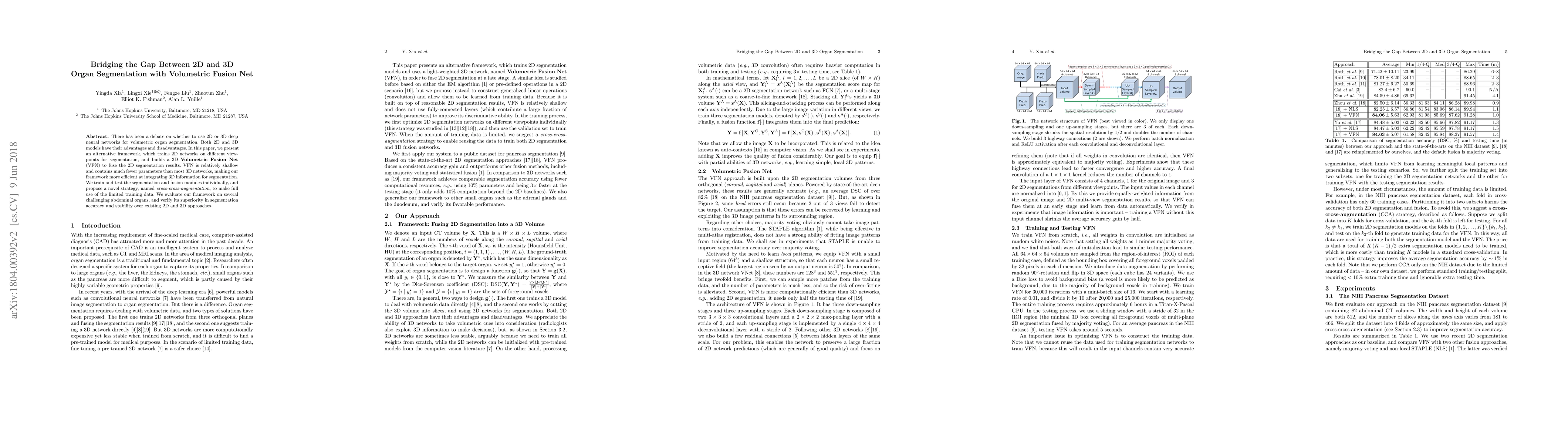
There has been a debate on whether to use 2D or 3D deep neural networks for volumetric organ segmentation. Both 2D and 3D models have their advantages and disadvantages. In this paper, we present an alternative framework, which trains 2D networks on different viewpoints for segmentation, and builds a 3D Volumetric Fusion Net (VFN) to fuse the 2D segmentation results. VFN is relatively shallow and contains much fewer parameters than most 3D networks, making our framework more efficient at integrating 3D information for segmentation. We train and test the segmentation and fusion modules individually, and propose a novel strategy, named cross-cross-augmentation, to make full use of the limited training data. We evaluate our framework on several challenging abdominal organs, and verify its superiority in segmentation accuracy and stability over existing 2D and 3D approaches.

AI Key Findings

Generated Sep 04, 2025

Methodology

The proposed approach combines 2D segmentation with a light-weighted 3D fusion module to improve organ segmentation in medical images.

Key Results

  • Improved 2D segmentation accuracy
  • Enhanced 3D fusion results
  • Reduced overfitting on limited training data

Significance

The proposed approach bridges the gap between 2D and 3D organ segmentation, enabling more accurate and reliable segmentation of challenging organs.

Technical Contribution

The proposed approach demonstrates a stable and efficient method for combining 2D and 3D organ segmentation, addressing the limitations of existing approaches.

Novelty

The use of light-weighted 3D fusion modules in 2D segmentation improves the accuracy and robustness of organ segmentation results, particularly on challenging organs.

Limitations

  • Limited to abdominal CT scans
  • Requires large amounts of annotated data for 3D fusion training

Future Work

  • Improving 3D fusion with more advanced architectures
  • Developing transfer learning methods for 2D segmentation
  • Applying the proposed approach to other medical imaging modalities

Paper Details

PDF Preview

Download PDF

Key Terms

segmentation (0.384)
fusion (0.229)
networks (0.202)
cross (0.182)
framework (0.180)
shallow (0.171)
builds (0.167)
make use (0.159)

Citation Network

Current paper (gray), citations (green), references (blue)

Display is limited for performance on very large graphs.

Current Paper
Citations
References
Click to view
Title Authors Year Actions

Comments (0)

This website uses cookies to ensure you get the best experience. Learn more