Few-Shot 3D Volumetric Segmentation with Multi-Surrogate Fusion
Publication
Metrics
AI Quick Summary
MSFSeg introduces a novel few-shot 3D volumetric segmentation framework that utilizes lightweight multi-surrogate fusion to accurately segment unseen 3D objects with minimal labeled data, demonstrating superior performance on benchmark datasets and challenging cross-domain segmentation tasks.
Paper Preview
Abstract
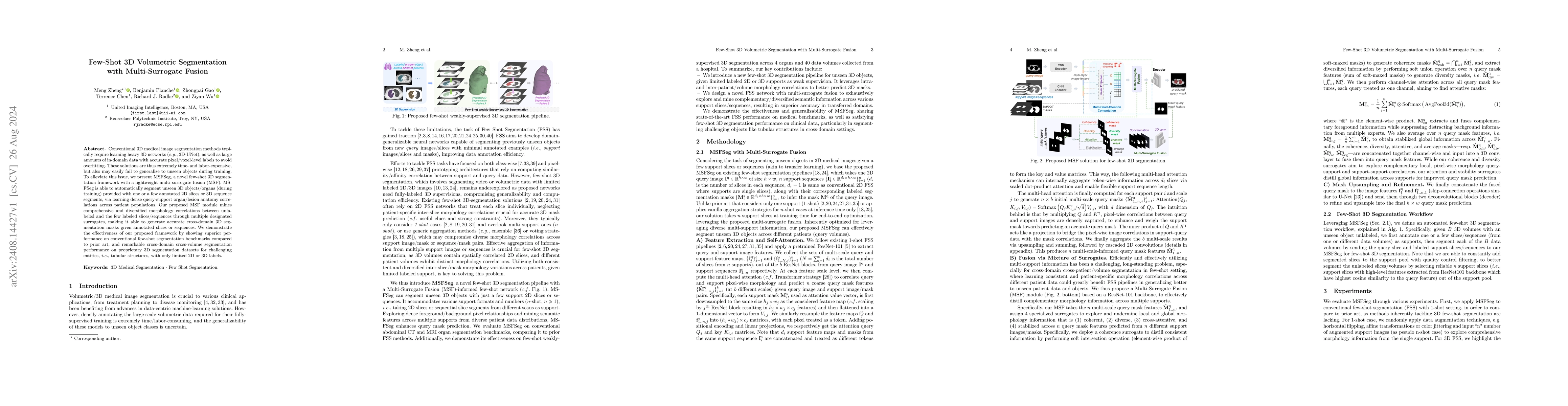
Conventional 3D medical image segmentation methods typically require learning heavy 3D networks (e.g., 3D-UNet), as well as large amounts of in-domain data with accurate pixel/voxel-level labels to avoid overfitting. These solutions are thus extremely time- and labor-expensive, but also may easily fail to generalize to unseen objects during training. To alleviate this issue, we present MSFSeg, a novel few-shot 3D segmentation framework with a lightweight multi-surrogate fusion (MSF). MSFSeg is able to automatically segment unseen 3D objects/organs (during training) provided with one or a few annotated 2D slices or 3D sequence segments, via learning dense query-support organ/lesion anatomy correlations across patient populations. Our proposed MSF module mines comprehensive and diversified morphology correlations between unlabeled and the few labeled slices/sequences through multiple designated surrogates, making it able to generate accurate cross-domain 3D segmentation masks given annotated slices or sequences. We demonstrate the effectiveness of our proposed framework by showing superior performance on conventional few-shot segmentation benchmarks compared to prior art, and remarkable cross-domain cross-volume segmentation performance on proprietary 3D segmentation datasets for challenging entities, i.e., tubular structures, with only limited 2D or 3D labels.
AI Key Findings
Get AI-generated insights about this paper's methodology, results, significance, and more — seven facets brought into focus.
Impact
Paper Details
Authors
PDF Preview
Citation Network
Current paper (gray), citations (green), references (blue)
Display is limited for performance on very large graphs.
Discussion 0