BriFiSeg: a deep learning-based method for semantic and instance segmentation of nuclei in brightfield images
Publication
Metrics
AI Quick Summary
This paper introduces BriFiSeg, a deep learning method for semantic and instance segmentation of nuclei in non-stained brightfield images, eliminating the need for specific sample preparation and staining. The study employs U-Net-based architectures and pre-trained encoders to achieve effective segmentation across diverse biological contexts.
Paper Preview
Abstract
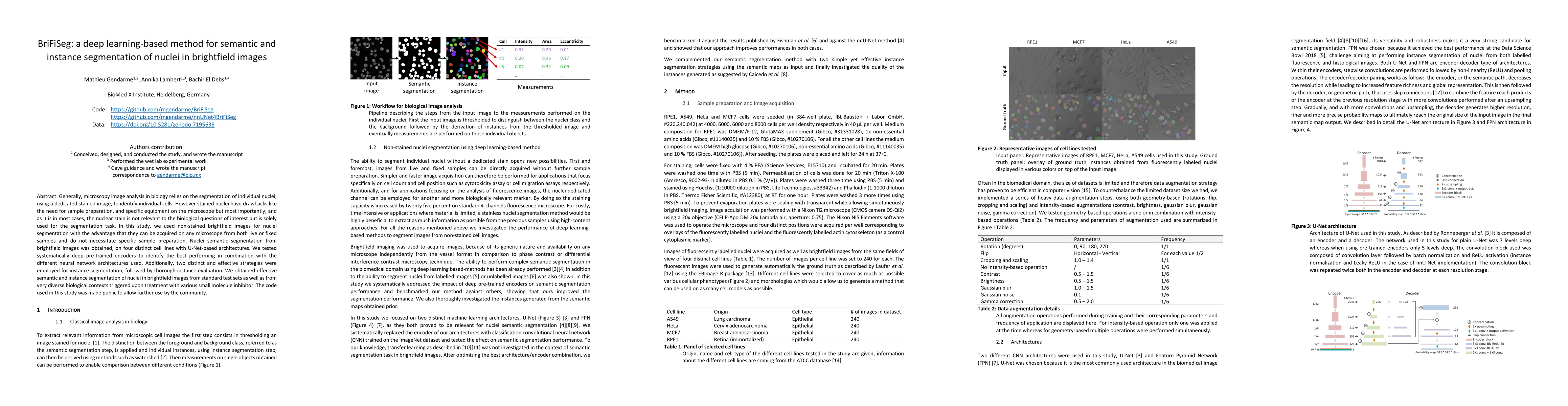
Generally, microscopy image analysis in biology relies on the segmentation of individual nuclei, using a dedicated stained image, to identify individual cells. However stained nuclei have drawbacks like the need for sample preparation, and specific equipment on the microscope but most importantly, and as it is in most cases, the nuclear stain is not relevant to the biological questions of interest but is solely used for the segmentation task. In this study, we used non-stained brightfield images for nuclei segmentation with the advantage that they can be acquired on any microscope from both live or fixed samples and do not necessitate specific sample preparation. Nuclei semantic segmentation from brightfield images was obtained, on four distinct cell lines with U-Net-based architectures. We tested systematically deep pre-trained encoders to identify the best performing in combination with the different neural network architectures used. Additionally, two distinct and effective strategies were employed for instance segmentation, followed by thorough instance evaluation. We obtained effective semantic and instance segmentation of nuclei in brightfield images from standard test sets as well as from very diverse biological contexts triggered upon treatment with various small molecule inhibitor. The code used in this study was made public to allow further use by the community.
AI Key Findings
Get AI-generated insights about this paper's methodology, results, significance, and more — seven facets brought into focus.
Impact
Paper Details
Authors
PDF Preview
Key Terms
Citation Network
Current paper (gray), citations (green), references (blue)
Display is limited for performance on very large graphs.
Discussion 0