ArxivLens

Quick Summary:

This paper demonstrates that StarDist, a deep learning method for nuclei segmentation, can effectively extend to histopathology images, achieving top results in the Colon Nuclei Identification and Counting (CoNIC) challenge 2022 for both segmentation and classification tasks. Experiments on the Lizard dataset substantiate its successful application.

DOI: 10.1109/ISBIC56247.2022.9854534
arXiv Id: 2203.02284
Date Published: 2022-08-22
Date Updated: 2022-08-22

Categories:

Key Terms (TF-IDF):

segmentation (0.388)
segmentation (0.368)
nuclei (0.368)
nuclei (0.366)
classification (0.244)
classification (0.242)
task (0.230)
task (0.225)
spot (0.223)
fluorescence (0.215)
conducting (0.206)
counting (0.199)
originally (0.198)
originally (0.197)
microscopy (0.197)
counting (0.195)
preliminary (0.189)
microscopy (0.187)
preliminary (0.182)
identification (0.173)
License: http://arxiv.org/licenses/nonexclusive-distrib/1.0/

Summary:

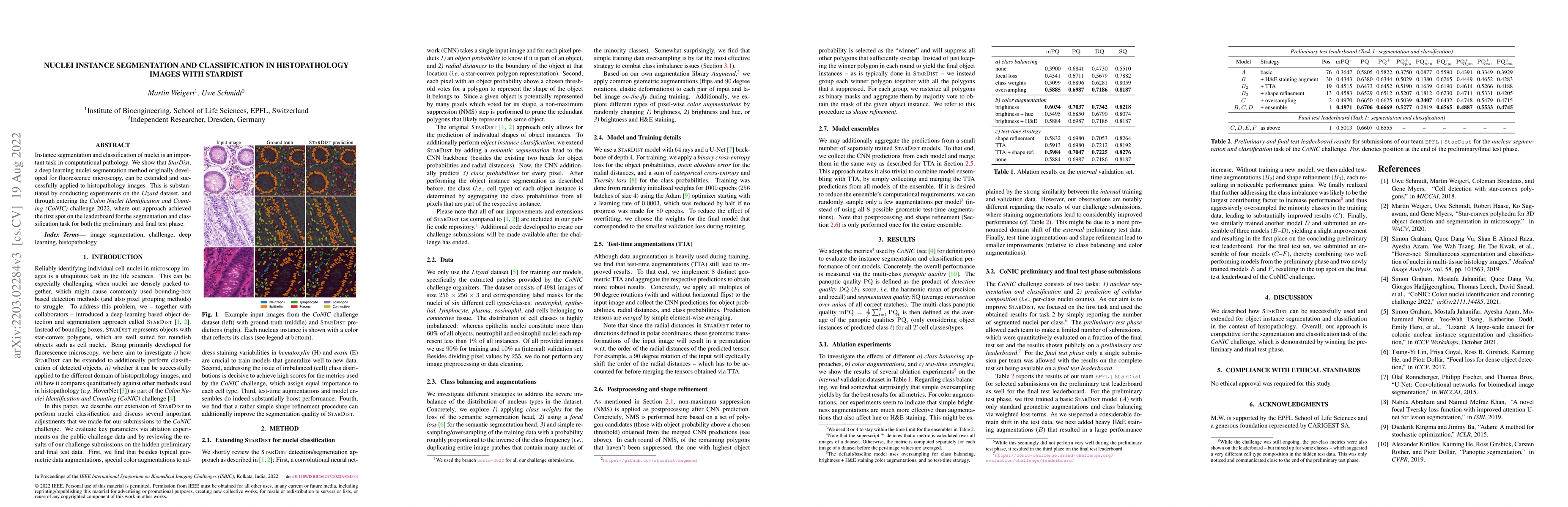
Instance segmentation and classification of nuclei is an important task in computational pathology. We show that StarDist, a deep learning nuclei segmentation method originally developed for fluorescence microscopy, can be extended and successfully applied to histopathology images. This is substantiated by conducting experiments on the Lizard dataset, and through entering the Colon Nuclei Identification and Counting (CoNIC) challenge 2022, where our approach achieved the first spot on the leaderboard for the segmentation and classification task for both the preliminary and final test phase.

Citation & Reference Network Visualization

This graph shows how this paper is connected to other research. The current paper is shown in dark gray, papers that cite it are in green, and papers it references are in blue. You can:

Current Paper
Papers citing this work (Citations)
Papers cited by this work (References)

• Hover over any node to see the paper title

• Click a node to view that paper's details

• Drag nodes to rearrange the graph

• Use the zoom controls (bottom right) to zoom in/out

Citations and References

Title Authors Year Actions
mTORC1 activation drives astrocyte reactivity in cortical tubers and brain organoid models of TSC
Thomas L. Li, John D. Blair, Taesun Yoo, Gerald A. Grant, Dirk Hockemeyer, Brenda E. Porter, Helen S. Bateup
2025
View Paper
A Lightweight and Extensible Cell Segmentation and Classification Model for Whole Slide Images
N. Shvetsov, T. Kilvaer, M. Tafavvoghi, Anders Sildnes, Kajsa Møllersen, Lill-ToveRasmussen Busund, L. A. Bongo
2025
View Paper
Loss-of-function mitochondrial DNA polymerase gamma variants cause vascular smooth muscle cells to secrete a diffusible mitogenic factor.
Samantha Rothwell, Irvin Ng, Sophia Shalchy-Tabrizi, Pola Kalinowski, Omnia M. Taha, Italia Paris, Angelica Baniqued, Lisa Lin, Michelle M. Mezei, Anna Lehman, Lisa M. Julian, Damon Poburko
2025
View Paper
Advancing antibiotic discovery with bacterial cytological profiling: a high-throughput solution to antimicrobial resistance
Jhonatan Salgado, James Rayner, Nikola Ojkic
2025
View Paper
SAMCell: Generalized Label-Free Biological Cell Segmentation with Segment Anything
Alexandra D. VandeLoo, Nathan J. Malta, Emilio Aponte, Caitlin van Zyl, Danfei Xu, Craig R. Forest
2025
View Paper
Sequence models for continuous cell cycle stage prediction from brightfield images
Louis-Alexandre Leger, Maxine Leonardi, Andrea Salati, Felix Naef, Martin Weigert
2025
View Paper
scCamAge: A context-aware prediction engine for cellular age, aging-associated bioactivities, and morphometrics.
Vishakha Gautam, Subhadeep Duari, Saveena Solanki, Mudit Gupta, Aayushi Mittal, Sakshi Arora, Anmol Aggarwal, Anmol Kumar Sharma, Sarthak Tyagi, Rathod Kunal Pankajbhai, Arushi Sharma, Sonam Chauhan, Shiva Satija, Suvendu Kumar, Sanjay Kumar Mohanty, Juhi Tayal, Nilesh Kumar Dixit, Debarka Sengupta, Anurag Mehta, Gaurav Ahuja
2025
View Paper
A Subtype Specific Probe for Targeted Magnetic Resonance Imaging of M2 Tumor-Associated Macrophages in Brain Tumors.
Yuancheng Li, Anbu Mozhi Thamizhchelvan, Hedi Ma, Jonathan Padelford, Zhaobin Zhang, Tianhe Wu, Quanquan Gu, Zi Wang, Hui Mao
2025
View Paper
An untargeted metabolomic study using MALDI-mass spectrometry imaging reveals region-specific biomarkers associated with bowel inflammation
Adele Ponzoni, Silvia Speca, Matthew Hartle, Amandine Gerstenberg, Aurore Tomezyk, Victor Senechal, S. Karnik, Laurent Dubuquoy, D. Launay, R. Deprez-Poulain, Mathieu Gaudin, Corinne Ramos, Benoit Deprez
2024
View Paper
Secondary metabolites of Vietnamese marine fungus Penicillium chermesinum 2104NT-1.3 and their cardioprotective activity
Ngo Thi Duy Ngoc, E. Yurchenko, Phan Thi Hoai Trinh, E. Menchinskaya, Trang Vo Thi Dieu, Anastasia D. Savagina, Artem Minin, P. D. Thinh, H. H. N. Khánh, Tran Thi Thanh Van, A. Yurchenko
2024
View Paper
Keep it accurate and robust: An enhanced nuclei analysis framework
Wenhua Zhang, Sen Yang, Meiwei Luo, Chuan He, Yuchen Li, Jun Zhang, Xiyue Wang, Fang Wang
2024
View Paper
How Good Are We? Evaluating Cell AI Foundation Models in Kidney Pathology with Human-in-the-Loop Enrichment
Junlin Guo, Siqi Lu, Can Cui, Ruining Deng, Tianyuan Yao, Zhewen Tao, Yizhe Lin, Marilyn Lionts, Quan Liu, Juming Xiong, Yu Wang, Shilin Zhao, Catie Chang, Mitchell Wilkes, Mengmeng Yin, Haichun Yang, Yuankai Huo
2024
View Paper
Cooperative motility emerges in crowds of T cells but not neutrophils
Inge M. N. Wortel, Jérémy Postat, Mihaela Mihaylova, Mauricio Merino, Aanya Bhagrath, Maryl Harris, Lin Wouters, Lucas Wiebke, Daniel R. Parisi, Judith N. Mandl, Johannes Textor
2024
View Paper
Vispro improves imaging analysis for Visium spatial transcriptomics
Huifang Ma, Yilong Qu, Anru R. Zhang, Zhicheng Ji
2024
View Paper
A Survey on Cell Nuclei Instance Segmentation and Classification: Leveraging Context and Attention
João D. Nunes, D. Montezuma, Domingos Oliveira, Tania Pereira, Jaime S. Cardoso
2024
View Paper
Real-time segmentation and classification of whole-slide images for tumor biomarker scoring
Md Jahid Hasan, Wan Siti Halimatul Munirah Wan Ahmad, Mohammad Faizal Ahmad Fauzi, J. T. H. Lee, S. Y. Khor, L. Looi, F. S. Abas, A. Adam, Elaine Wan Ling Chan
2024
View Paper
NuLite - Lightweight and Fast Model for Nuclei Instance Segmentation and Classification
C. Tommasino, Cristiano Russo, A. M. Rinaldi
2024
View Paper
A Survey on Cell Nuclei Instance Segmentation and Classification: Leveraging Context and Attention
João D. Nunes, D. Montezuma, Domingos Oliveira, Tania Pereira, Jaime S. Cardoso
2024
View Paper
PFPs: Prompt-guided Flexible Pathological Segmentation for Diverse Potential Outcomes Using Large Vision and Language Models
Can Cui, Ruining Deng, Junlin Guo, Quan Liu, Tianyuan Yao, Haichun Yang, Yuankai Huo
2024
View Paper
SR-CACO-2: A Dataset for Confocal Fluorescence Microscopy Image Super-Resolution
Soufiane Belharbi, Mara K M Whitford, Phuong Hoang, Shakeeb Murtaza, Luke McCaffrey, Eric Granger
2024
View Paper
The development of artificial intelligence in the histological diagnosis of Inflammatory Bowel Disease (IBD-AI).
Cesare Furlanello, Nicole Bussola, Nicolò Merzi, Giovanni Pievani Trapletti, M. Cadei, R. Del Sordo, Angelo Sidoni, C. Ricci, F. Lanzarotto, T. Parigi, Vincenzo Villanacci
2024
View Paper
Integrating Spatial and Morphological Characteristics into Melanoma Prognosis: A Computational Approach
Chang Bian, Garry Ashton, Megan Grant, Valeria Pavet Rodriguez, Isabel Peset Martin, A. Tsakiroglou, Martin Cook, Martin Fergie
2024
View Paper
Quantification of collective signalling in time-lapse microscopy images
M. Dobrzyński, Benjamin Grädel, P. Gagliardi, Olivier Pertz
2024
View Paper
Pseudo-spectral angle mapping to improve immune cell classification in highly multiplexed fluorescence microscopy images
Madeleine S. Durkee, Junting Ai, Gabriel Casella, Thao Cao, Marcus R. Clark, Maryellen L. Giger
2024
View Paper
Combining Datasets with Different Label Sets for Improved Nucleus Segmentation and Classification
Amruta Parulekar, Utkarsh Kanwat, Ravi Kant Gupta, Medha Chippa, Thomas Jacob, Tripti Bameta, Swapnil Rane, Amit Sethi
2023
View Paper
H&E image analysis pipeline for quantifying morphological features
V. Ariotta, Oskari Lehtonen, S. Salloum, Giulia Micoli, K. Lavikka, Ville Rantanen, J. Hynninen, A. Virtanen, S. Hautaniemi
2023
View Paper
Image Analysis of Nuclei Histopathology Using Deep Learning: A Review of Segmentation, Detection, and Classification
Mukul Kadaskar, Nagamma Patil
2023
View Paper
CellViT: Vision Transformers for Precise Cell Segmentation and Classification
Fabian Hörst, Moritz Rempe, Lukas Heine, Constantin Seibold, J. Keyl, G. Baldini, S. Ugurel, J. Siveke, Barbara Grünwald, J. Egger, J. Kleesiek
2023
View Paper
Predicting cell type counts in whole slide histology images using evidential multi-task learning
N. R. Gudhe, M. Sudah, A. Mannermaa, V. Kosma, Hamid Behravan
2023
View Paper
High Expression of IRS-1, RUNX3 and SMAD4 Are Positive Prognostic Factors in Stage I–III Colon Cancer
Hallgeir Selven, L. Busund, S. Andersen, M. Pedersen, A. Lombardi, T. Kilvaer
2023
View Paper
Annotating for Artificial Intelligence Applications in Digital Pathology: A Practical Guide for Pathologists and Researchers.
D. Montezuma, Sara P. Oliveira, Pedro C. Neto, Domingos Oliveira, Ana Monteiro, J. S. Cardoso, I. Macedo-Pinto
2023
View Paper
Ciscnet - a Single-Branch Cell Nucleus Instance Segmentation and Classification Network
Moritz Böhland, Oliver Neumann, Marcel P. Schilling, M. Reischl, R. Mikut, Katharina Löffler, Tim Scherr
2022
View Paper

Comments (0)

This website uses cookies to ensure you get the best experience. Learn more