ArxivLens
M
Researcher

Martin Weigert

8 papers on arXiv

8 arXiv Papers
17 Total Publications
Profile

Academic Profile

Name Martin Weigert
Total Papers 17
Scholar Profile View on Semantic Scholar
Metrics

Statistics

8 arXiv Papers
17 Total Publications
Publications

Papers on arXiv

Thumbnail for Trackastra: Transformer-based cell tracking for live-cell microscopy
arXiv
Trackastra: Transformer-based cell tracking for live-cell microscopy

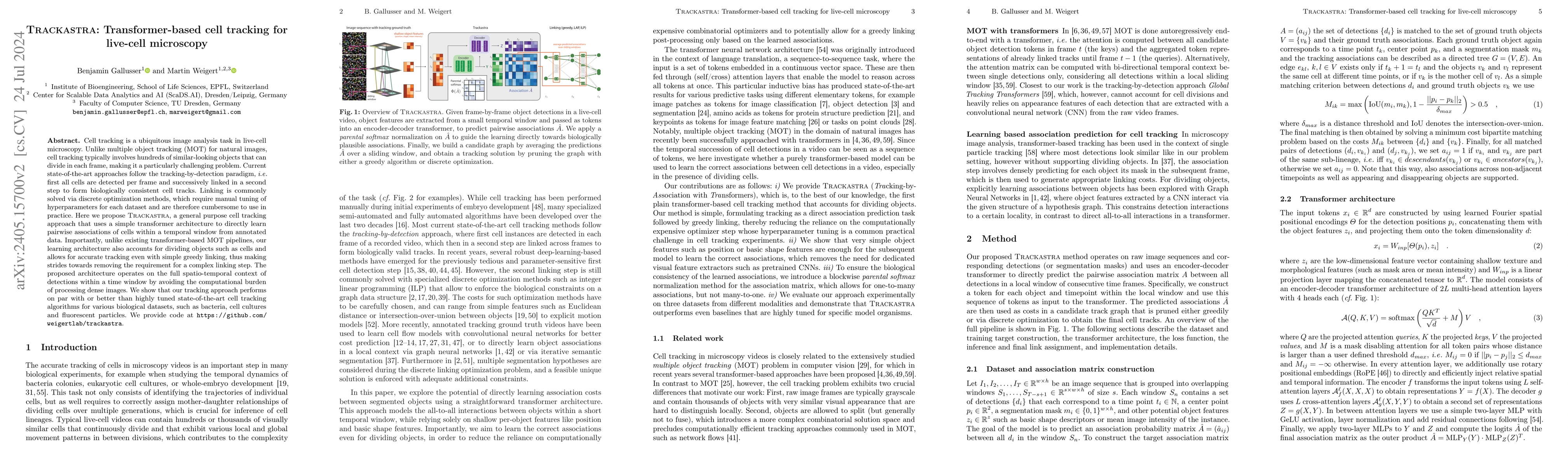
Cell tracking is an omnipresent image analysis task in live-cell microscopy. It is similar to multiple object tracking (MOT), however, each frame contains hundreds of similar-looking objects that ca...

Thumbnail for Self-supervised dense representation learning for live-cell microscopy
  with time arrow prediction
arXiv
Self-supervised dense representation learning for live-cell microscopy with time arrow prediction

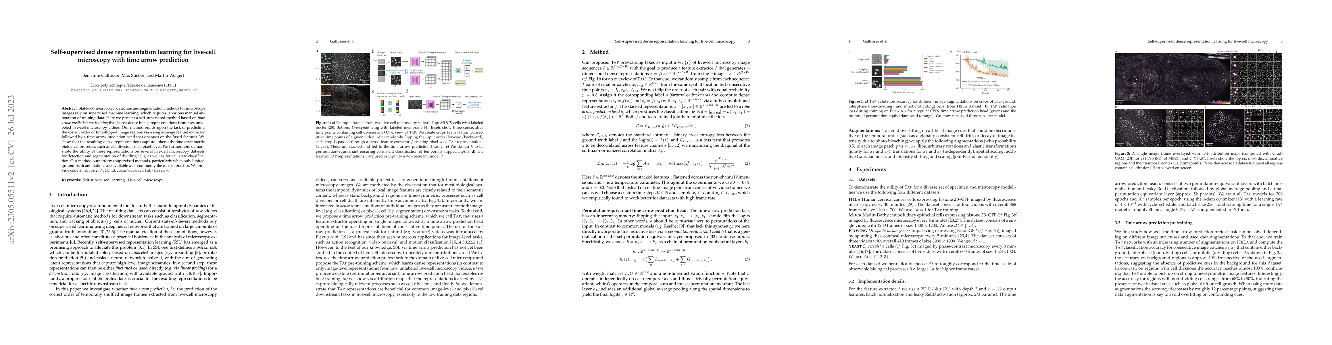
State-of-the-art object detection and segmentation methods for microscopy images rely on supervised machine learning, which requires laborious manual annotation of training data. Here we present a s...

Thumbnail for CoNIC Challenge: Pushing the Frontiers of Nuclear Detection,
  Segmentation, Classification and Counting
arXiv
CoNIC Challenge: Pushing the Frontiers of Nuclear Detection, Segmentation, Classification and Counting

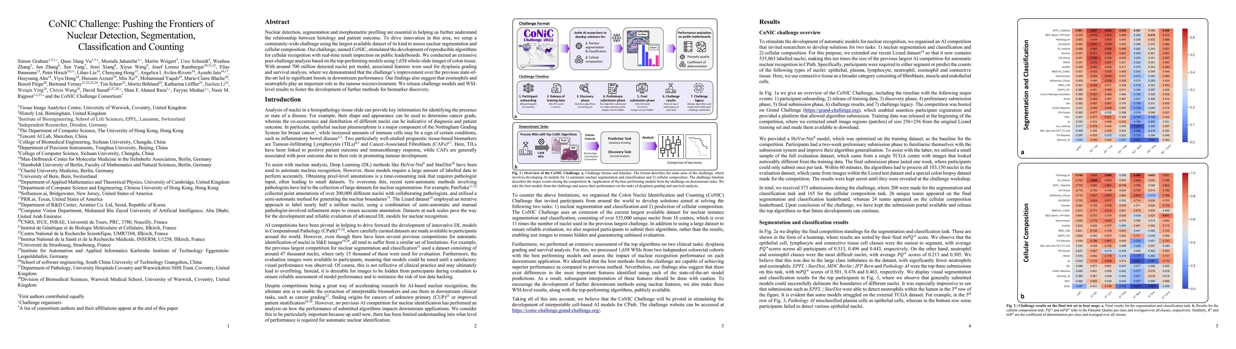
Nuclear detection, segmentation and morphometric profiling are essential in helping us further understand the relationship between histology and patient outcome. To drive innovation in this area, we...

Thumbnail for Organelle-specific segmentation, spatial analysis, and visualization of
  volume electron microscopy datasets
arXiv
Organelle-specific segmentation, spatial analysis, and visualization of volume electron microscopy datasets

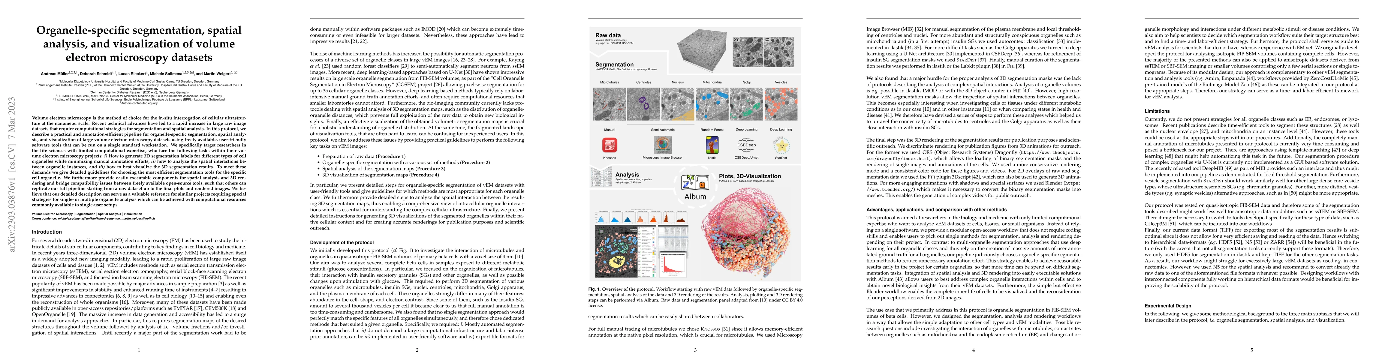
Volume electron microscopy is the method of choice for the in-situ interrogation of cellular ultrastructure at the nanometer scale. Recent technical advances have led to a rapid increase in large ra...

Thumbnail for Nuclei instance segmentation and classification in histopathology images
  with StarDist
arXiv
Nuclei instance segmentation and classification in histopathology images with StarDist

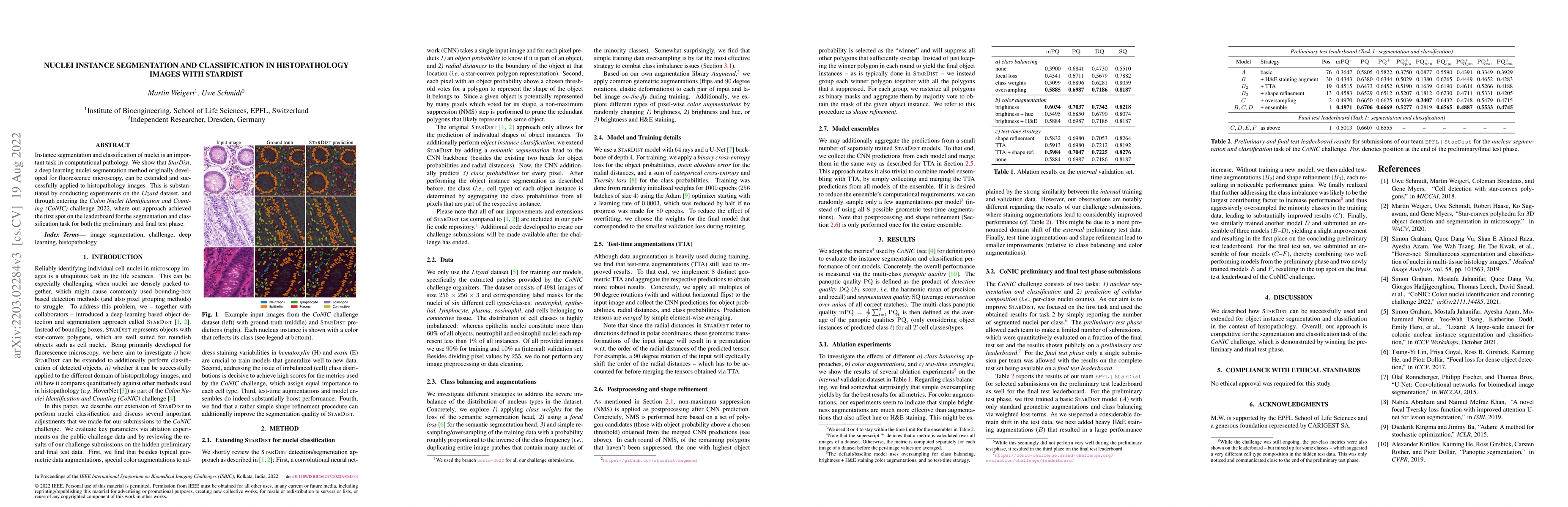
Instance segmentation and classification of nuclei is an important task in computational pathology. We show that StarDist, a deep learning nuclei segmentation method originally developed for fluores...

Thumbnail for An interpretable automated detection system for FISH-based HER2 oncogene
  amplification testing in histo-pathological routine images of breast and
  gastric cancer diagnostics
arXiv
An interpretable automated detection system for FISH-based HER2 oncogene amplification testing in histo-pathological routine images of breast and gastric cancer diagnostics

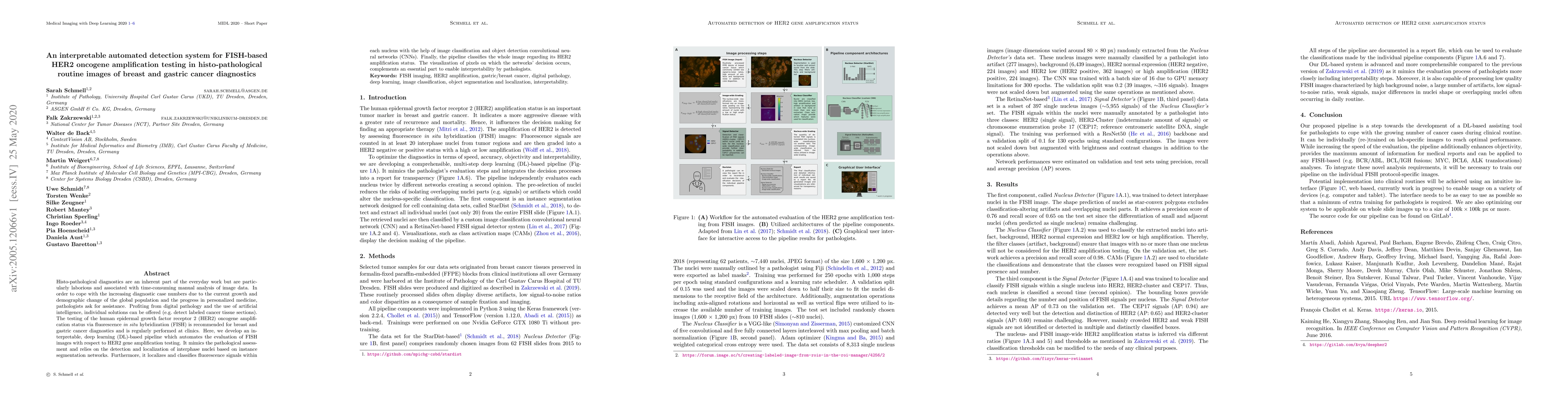
Histo-pathological diagnostics are an inherent part of the everyday work but are particularly laborious and associated with time-consuming manual analysis of image data. In order to cope with the in...

Thumbnail for Sequence models for continuous cell cycle stage prediction from
  brightfield images
arXiv
Sequence models for continuous cell cycle stage prediction from brightfield images

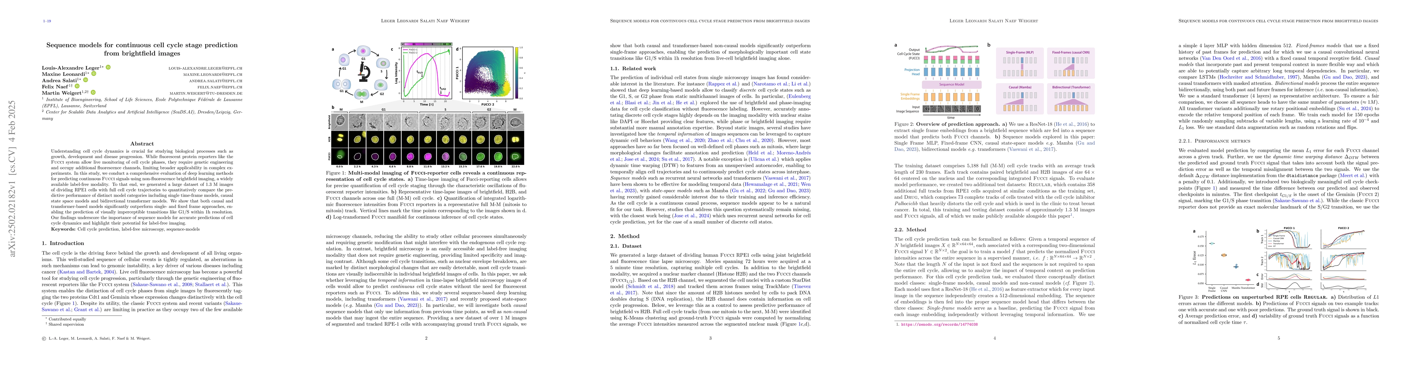
Understanding cell cycle dynamics is crucial for studying biological processes such as growth, development and disease progression. While fluorescent protein reporters like the Fucci system allow live...

Thumbnail for MuViT: Multi-Resolution Vision Transformers for Learning Across Scales in Microscopy
arXiv
MuViT: Multi-Resolution Vision Transformers for Learning Across Scales in Microscopy

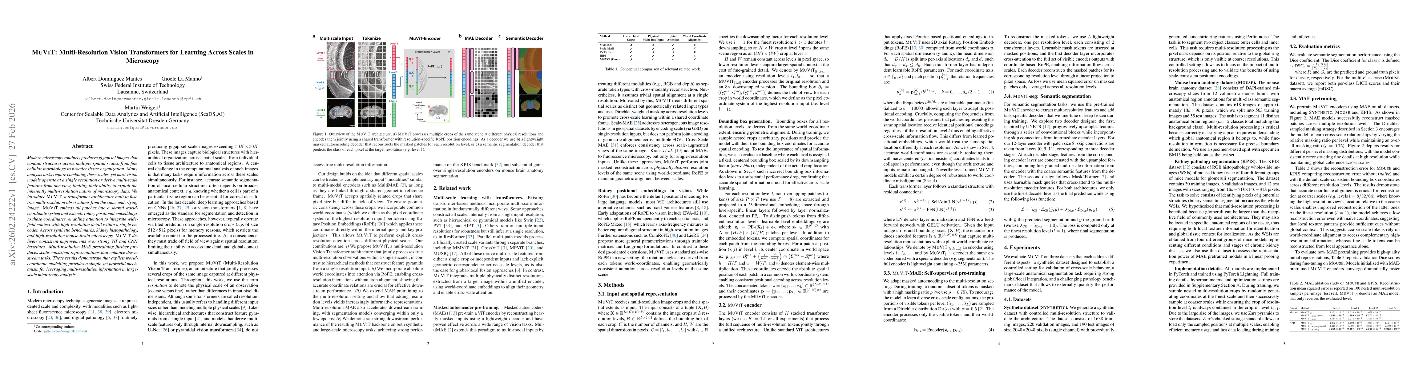
Modern microscopy routinely produces gigapixel images that contain structures across multiple spatial scales, from fine cellular morphology to broader tissue organization. Many analysis tasks require ...

This website uses cookies to ensure you get the best experience. Learn more