This paper presents a user-friendly computational pipeline for organelle-specific segmentation, spatial analysis, and visualization of large volume electron microscopy datasets, targeting life scientists with limited computational expertise. The protocol details efficient segmentation tools, spatial interaction analysis, and 3D rendering using freely available software, enabling replication of the workflow from raw datasets to final visualizations.
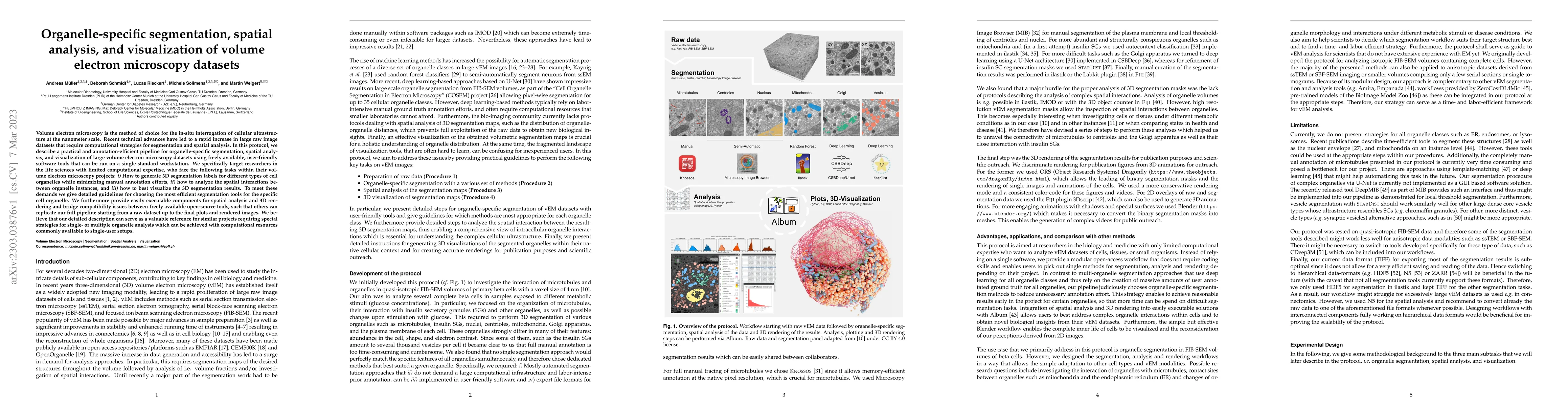
Volume electron microscopy is the method of choice for the in-situ interrogation of cellular ultrastructure at the nanometer scale. Recent technical advances have led to a rapid increase in large raw image datasets that require computational strategies for segmentation and spatial analysis. In this protocol, we describe a practical and annotation-efficient pipeline for organelle-specific segmentation, spatial analysis, and visualization of large volume electron microscopy datasets using freely available, user-friendly software tools that can be run on a single standard workstation. We specifically target researchers in the life sciences with limited computational expertise, who face the following tasks within their volume electron microscopy projects: i) How to generate 3D segmentation labels for different types of cell organelles while minimizing manual annotation efforts, ii) how to analyze the spatial interactions between organelle instances, and iii) how to best visualize the 3D segmentation results. To meet these demands we give detailed guidelines for choosing the most efficient segmentation tools for the specific cell organelle. We furthermore provide easily executable components for spatial analysis and 3D rendering and bridge compatibility issues between freely available open-source tools, such that others can replicate our full pipeline starting from a raw dataset up to the final plots and rendered images. We believe that our detailed description can serve as a valuable reference for similar projects requiring special strategies for single- or multiple organelle analysis which can be achieved with computational resources commonly available to single-user setups.
This graph shows how this paper is connected to other research. The current paper is shown in dark gray, papers that cite it are in green, and papers it references are in blue. You can:
• Hover over any node to see the paper title
• Click a node to view that paper's details
• Drag nodes to rearrange the graph
• Use the zoom controls (bottom right) to zoom in/out
Title | Authors | Year | Actions |
---|---|---|---|
Structure, interaction and nervous connectivity of beta cell primary cilia
|
A. Müller, Nikolai Klena, Song Pang, Leticia Elizabeth, Galicia Garcia, Davud Sulaymankhil, Oleksandra Topcheva, Monika Seliskar, Hassan Mziaut, E. Schöniger, Daniela Friedland, N. Kipke, S. Kretschmar, C. Münster, Jürgen Weitz, Marius Distler, Thomas Kurth, Deborah Schmidt, Harald F. Hess, Shan Xu, Gaia Pigino, M. Solimena
|
2024
|
View Paper |
Nellie: automated organelle segmentation, tracking and hierarchical feature extraction in 2D/3D live-cell microscopy
|
Austin E. Y. T. Lefebvre, Gabriel Sturm, Ting-Yu Lin, Emily Stoops, Magdalena Preciado Lopez, Benjamin Kaufmann-Malaga, Kayley Hake
|
2024
|
View Paper |
Edinburgh Research Explorer Evolution and Impact of High Content Imaging
|
Gregory P. Way, Slas Discovery, Heba Sailem, S. Shave, Richard Kasprowicz, N. Carragher
|
View Paper |
Title | Authors | Year | Actions |
---|---|---|---|
Instance segmentation of mitochondria in electron microscopy images with a generalist deep learning model trained on a diverse dataset.
|
Ryan Conrad, Kedar Narayan
|
2023
|
View Paper |
Integration of ER protein quality control mechanisms defines β cell function and ER architecture
|
N. Shrestha, M. Torres, Jason Zhang, You Lu, L. Haataja, Rachel B. Reinert, J. Knupp, Yu-Jie Chen, Gunes Parlakgul, A. P. Arruda, Billy Tsai, P. Arvan, Ling Qi
|
2022
|
View Paper |
A serotonergic axon-cilium synapse drives nuclear signaling to alter chromatin accessibility
|
S. Sheu, S. Upadhyayula, V. Dupuy, Song Pang, Fei Deng, Jinxia Wan, D. Walpita, H. Pasolli, Justin Houser, Silvia Sánchez-Martínez, S. Brauchi, Sambashiva Banala, Melanie Freeman, C. Xu, T. Kirchhausen, H. Hess, L. Lavis, Yulong Li, S. Chaumont-Dubel, D. Clapham
|
2022
|
View Paper |
Weighted average ensemble-based semantic segmentation in biological electron microscopy images
|
Kavitha Shaga Devan, H. Kestler, C. Read, P. Walther
|
2022
|
View Paper |
Deep neural network automated segmentation of cellular structures in volume electron microscopy
|
Benjamin Gallusser, G. Maltese, G. Di Caprio, T. Vadakkan, Anwesha Sanyal, Elliott Somerville, M. Sahasrabudhe, Justin O'Connor, Martin Weigert, T. Kirchhausen
|
2022
|
View Paper |
Volume electron microscopy
|
C. Peddie, C. Genoud, A. Kreshuk, Kimberly I. Meechan, Kristina D. Micheva, Kedar Narayan, Constantin Pape, R. Parton, N. Schieber, Y. Schwab, B. Titze, P. Verkade, Aubrey V. Weigel, L. Collinson
|
2022
|
View Paper |
BioImage Model Zoo: A Community-Driven Resource for Accessible Deep Learning in BioImage Analysis
|
Ouyang Wei, Fynn Beuttenmueller, Estibaliz Gómez-de-Mariscal, Constantin Pape, Tom Burke, C. García-López-de-Haro, Craig T Russell, Lucía Moya-Sans, Cristina de-la-Torre-Gutiérrez, Deborah Schmidt, D. Kutra, Maksim Novikov, Martin Weigert, Uwe Schmidt, P. Bankhead, Guillaume Jacquemet, D. Sage, Ricardo Henriques, A. Muñoz-Barrutia, E. Lundberg, Florian Jug, A. Kreshuk
|
2022
|
View Paper |
Regulation of liver subcellular architecture controls metabolic homeostasis
|
Güneş Parlakgül, A. P. Arruda, Song Pang, Erika Cagampan, Nina Min, Ekin Güney, G. Y. Lee, K. Inouye, H. Hess, C. Xu, G. Hotamışlıgil
|
2022
|
View Paper |
Profiling cellular diversity in sponges informs animal cell type and nervous system evolution
|
J. Musser, K. Schippers, M. Nickel, Giulia Mizzon, A. Kohn, Constantin Pape, P. Ronchi, Nikolaos Papadopoulos, Alexander J. Tarashansky, J. Hammel, F. Wolf, Cong Liang, Ana Hernández-Plaza, C. Cantalapiedra, K. Achim, N. Schieber, Leslie Pan, Fabian Ruperti, W. Francis, S. Vargas, Svenja Kling, Maike Renkert, M. Polikarpov, G. Bourenkov, Roberto Feuda, Imre Gáspár, P. Burkhardt, Bo Wang, P. Bork, Martin Beck, T. Schneider, A. Kreshuk, G. Wörheide, J. Huerta-Cepas, Y. Schwab, L. Moroz, D. Arendt
|
2021
|
View Paper |
Labkit: Labeling and Segmentation Toolkit for Big Image Data
|
Matthias Arzt, Joran Deschamps, C. Schmied, T. Pietzsch, Deborah Schmidt, R. Haase, Florian Jug
|
2021
|
View Paper |
Whole-cell organelle segmentation in volume electron microscopy
|
Larissa Heinrich, Davis Bennett, David Ackerman, Woohyun Park, J. Bogovic, N. Eckstein, A. Petruncio, Jody Clements, Song Pang, Shan Xu, Jan Funke, Wyatt L. Korff, H. Hess, J. Lippincott-Schwartz, S. Saalfeld, Aubrey V. Weigel, Project Team, Riasat Ali, Rebecca Arruda, Rohit Bahtra, Destiny Nguyen
|
2021
|
View Paper |
An open-access volume electron microscopy atlas of whole cells and tissues
|
C. Xu, Song Pang, G. Shtengel, A. Müller, A. Ritter, Huxley K. Hoffman, Shin-ya Takemura, Zhiyuan Lu, H. Pasolli, N. Iyer, Jeeyun Chung, Davis Bennett, Aubrey V. Weigel, Melanie Freeman, Schuyler B. van Engelenburg, Tobias C. Walther, R. V. Farese, Jennifer Lippincott-Schwartz, Ira Mellman, M. Solimena, H. Hess
|
2021
|
View Paper |
Album: a framework for scientific data processing with software solutions of heterogeneous tools
|
J. Albrecht, D. Schmidt, Kyle Harrington
|
2021
|
View Paper |
Deep learning for bioimage analysis
|
Adrien Hallou, Hannah G. Yevick, Bianca Dumitrascu, V. Uhlmann
|
2021
|
View Paper |
Automatic detection of synaptic partners in a whole-brain Drosophila electron microscopy data set
|
Julia M. Buhmann, Arlo Sheridan, Caroline Malin-Mayor, P. Schlegel, S. Gerhard, Tom Kazimiers, R. Krause, Tri M. Nguyen, Larissa Heinrich, W. Lee, Rachel I. Wilson, S. Saalfeld, G. Jefferis, D. Bock, Srinivas C. Turaga, Matthew Cook, Jan Funke
|
2021
|
View Paper |
Deep learning for automatic segmentation of the nuclear envelope in electron microscopy data, trained with volunteer segmentations
|
Helen Spiers, Harry Songhurst, Luke Nightingale, J. de Folter, R. Hutchings, C. Peddie, A. Weston, A. Strange, S. Hindmarsh, C. Lintott, L. Collinson, Martin L. Jones
|
2021
|
View Paper |
Democratising deep learning for microscopy with ZeroCostDL4Mic
|
Lucas von Chamier, Romain F. Laine, Johanna Jukkala, Christoph Spahn, Daniel Krentzel, E. Nehme, Martina Lerche, Sara Hernández-Pérez, P. Mattila, Eleni Karinou, S. Holden, A. Solak, Alexander Krull, T. Buchholz, Martin L. Jones, Loic A. Royer, Christophe Leterrier, Y. Shechtman, Florian Jug, M. Heilemann, Guillaume Jacquemet, Ricardo Henriques
|
2021
|
View Paper |
ER-to-Golgi protein delivery through an interwoven, tubular network extending from ER
|
Aubrey V. Weigel, Chi-Lun Chang, G. Shtengel, C. Xu, D. P. Hoffman, Melanie Freeman, N. Iyer, J. Aaron, S. Khuon, J. Bogovic, Wei-Lin Qiu, H. Hess, J. Lippincott-Schwartz
|
2021
|
View Paper |
Morphological bases of phytoplankton energy management and physiological responses unveiled by 3D subcellular imaging
|
Clarisse Uwizeye, J. Decelle, P. Jouneau, Serena Flori, B. Gallet, Jean-Baptiste Keck, D. Bo, C. Moriscot, C. Seydoux, F. Chevalier, N. Schieber, R. Templin, Guillaume Allorent, Florence Courtois, G. Curien, Y. Schwab, G. Schoehn, S. Zeeman, D. Falconet, G. Finazzi
|
2021
|
View Paper |
Local shape descriptors for neuron segmentation
|
Arlo Sheridan, Tri M. Nguyen, Diptodip Deb, W. Lee, S. Saalfeld, Srinivas C. Turaga, U. Manor, Jan Funke
|
2021
|
View Paper |
3D FIB-SEM reconstruction of microtubule–organelle interaction in whole primary mouse β cells
|
A. Müller, Deborah Schmidt, C. Xu, Song Pang, Joyson Verner D’Costa, S. Kretschmar, C. Münster, T. Kurth, Florian Jug, Martin Weigert, Harald F. Hess, M. Solimena
|
2020
|
View Paper |
CEM500K, a large-scale heterogeneous unlabeled cellular electron microscopy image dataset for deep learning
|
Ryan Conrad, Kedar Narayan
|
2020
|
View Paper |
SplineDist: Automated Cell Segmentation with Spline Curves
|
Soham Mandal, V. Uhlmann
|
2020
|
View Paper |
Microtubule Tracking in Electron Microscopy Volumes
|
N. Eckstein, Julia M. Buhmann, Matthew Cook, Jan Funke
|
2020
|
View Paper |
DeepMIB: User-friendly and open-source software for training of deep learning network for biological image segmentation
|
I. Belevich, E. Jokitalo
|
2020
|
View Paper |
Array programming with NumPy
|
Charles R. Harris, K. Millman, S. Walt, R. Gommers, Pauli Virtanen, D. Cournapeau, Eric Wieser, Julian Taylor, Sebastian Berg, Nathaniel J. Smith, Robert Kern, Matti Picus, Stephan Hoyer, M. Kerkwijk, M. Brett, A. Haldane, Jaime Fern'andez del R'io, Marcy Wiebe, Pearu Peterson, Pierre G'erard-Marchant, Kevin Sheppard, Tyler Reddy, Warren Weckesser, Hameer Abbasi, C. Gohlke, T. Oliphant
|
2020
|
View Paper |
A connectome and analysis of the adult Drosophila central brain
|
Louis K. Scheffer, C. Xu, M. Januszewski, Zhiyuan Lu, Zhiyuan Lu, Shin-ya Takemura, K. Hayworth, Gary B. Huang, K. Shinomiya, Jeremy Maitlin-Shepard, S. Berg, Jody Clements, Philip M. Hubbard, W. Katz, L. Umayam, Ting Zhao, David Ackerman, T. Blakely, J. Bogovic, Tom Dolafi, Dagmar Kainmueller, Takashi Kawase, Khaled Khairy, Laramie Leavitt, Peter H. Li, Larry F. Lindsey, Nicole L. Neubarth, D. J. Olbris, H. Otsuna, Eric T. Trautman, Masayoshi Ito, Masayoshi Ito, A. S. Bates, Jens Goldammer, Jens Goldammer, T. Wolff, Rob Svirskas, P. Schlegel, Erika Neace, Christopher Knecht, Chelsea X. Alvarado, Dennis Bailey, Samantha Ballinger, J. Borycz, Brandon S Canino, Natasha Cheatham, Michael Cook, M. Dreher, O. Duclos, Bryon Eubanks, K. Fairbanks, Samantha Finley, N. Forknall, Audrey Francis, G. Hopkins, Emily M Joyce, SungJin Kim, Nicole Kirk, Julie Kovalyak, Shirley A Lauchie, Alanna Lohff, Charli A Maldonado, Emily A Manley, Sari McLin, Caroline Mooney, Miatta Ndama, Omotara Ogundeyi, Nneoma Okeoma, Christopher Ordish, Nicholas Padilla, Christopher Patrick, Tyler Paterson, Elliott Phillips, E. M. Phillips, Neha Rampally, C. Ribeiro, Madelaine K Robertson, J. Rymer, S. Ryan, Megan Sammons, Anne K. Scott, Ashley L Scott, A. Shinomiya, Claire Smith, Kelsey Smith, Natalie Smith, Margaret A. Sobeski, A. Suleiman, Jackie Swift, Satoko Takemura, Iris Talebi, Dorota Tarnogorska, Emily Tenshaw, Temour Tokhi, J. Walsh, Tansy Yang, J. Horne, Feng Li, Ruchi Parekh, P. Rivlin, V. Jayaraman, M. Costa, G. Jefferis, G. Jefferis, Kei Ito, Kei Ito, Kei Ito, S. Saalfeld, R. George, I. Meinertzhagen, I. Meinertzhagen, G. Rubin, H. Hess, Viren Jain, Stephen M. Plaza
|
2020
|
View Paper |
Whole-body integration of gene expression and single-cell morphology
|
H. Vergara, Constantin Pape, Kimberly I. Meechan, Valentyna Zinchenko, C. Genoud, A. Wanner, B. Titze, R. Templin, Paola Y. Bertucci, O. Simakov, P. Machado, Emily Savage, Y. Schwab, R. Friedrich, A. Kreshuk, C. Tischer, D. Arendt
|
2020
|
View Paper |
An interactive ImageJ plugin for semi-automated image denoising in electron microscopy
|
J. Roels, F. Vernaillen, A. Kremer, Amanda Gonçalves, J. Aelterman, H. Luong, B. Goossens, W. Philips, S. Lippens, Y. Saeys
|
2020
|
View Paper |
AMST: Alignment to Median Smoothed Template for Focused Ion Beam Scanning Electron Microscopy Image Stacks
|
Julian Hennies, José Miguel Serra Lleti, N. Schieber, R. Templin, A. Steyer, Y. Schwab
|
2020
|
View Paper |
Cellpose: a generalist algorithm for cellular segmentation
|
C. Stringer, Tim Wang, Michalis Michaelos, Marius Pachitariu
|
2020
|
View Paper |
Reconstruction of motor control circuits in adult Drosophila using automated transmission electron microscopy
|
Jasper T Maniates-Selvin, David Grant Colburn Hildebrand, Brett J. Graham, Aaron T. Kuan, Logan A. Thomas, Tri M. Nguyen, Julia M. Buhmann, Anthony W. Azevedo, Brendan L. Shanny, Jan Funke, John C. Tuthill, W. Lee
|
2020
|
View Paper |
You Can
|
Meg Busing
|
2019
|
View Paper |
A petascale automated imaging pipeline for mapping neuronal circuits with high-throughput transmission electron microscopy
|
W. Yin, D. Brittain, J. Borseth, Marie E. Scott, Derric Williams, Jed Perkins, C. Own, M. Murfitt, R. Torres, D. Kapner, Adam A. Bleckert, D. Castelli, David Reid, W. Lee, Brett J. Graham, Marc M. Takeno, D. Bumbarger, Colin Farrell, R. Reid, N. D. da Costa
|
2019
|
View Paper |
ilastik: interactive machine learning for (bio)image analysis
|
S. Berg, D. Kutra, Thorben Kroeger, C. Straehle, Bernhard X. Kausler, Carsten Haubold, Martin Schiegg, J. Ales, T. Beier, Markus Rudy, Kemal Eren, Jaime I Cervantes, Buote Xu, Fynn Beuttenmueller, A. Wolny, Chong Zhang, U. Köthe, F. Hamprecht, A. Kreshuk
|
2019
|
View Paper |
Correlative three-dimensional super-resolution and block-face electron microscopy of whole vitreously frozen cells
|
D. P. Hoffman, G. Shtengel, Cangshan Xu, K. Campbell, Melanie Freeman, Lei Wang, D. Milkie, H. Pasolli, N. Iyer, J. Bogovic, D. Stabley, A. Shirinifard, Song Pang, D. Peale, K. Schaefer, W. Pomp, Chi-Lun Chang, J. Lippincott-Schwartz, T. Kirchhausen, D. Solecki, E. Betzig, H. Hess
|
2019
|
View Paper |
Profiling cellular diversity in sponges informs animal cell type and nervous system evolution
|
J. Musser, K. Schippers, M. Nickel, Giulia Mizzon, A. Kohn, Constantin Pape, J. Hammel, Florian Wolf, Cong Liang, Ana Hernández-Plaza, K. Achim, N. Schieber, W. Francis, Sergio Vargas R., Svenja Kling, Maike Renkert, Roberto Feuda, Imre Gáspár, P. Burkhardt, P. Bork, Martin Beck, A. Kreshuk, G. Wörheide, J. Huerta-Cepas, Y. Schwab, L. Moroz, D. Arendt
|
2019
|
View Paper |
Star-convex Polyhedra for 3D Object Detection and Segmentation in Microscopy
|
Martin Weigert, Uwe Schmidt, R. Haase, Ko Sugawara, E. Myers
|
2019
|
View Paper |
SciPy 1.0: fundamental algorithms for scientific computing in Python
|
Pauli Virtanen, R. Gommers, T. Oliphant, Matt Haberland, Tyler Reddy, D. Cournapeau, Evgeni Burovski, Pearu Peterson, Warren Weckesser, Jonathan Bright, S. J. van der Walt, M. Brett, Joshua Wilson, K. Millman, N. Mayorov, Andrew R. J. Nelson, E. Jones, Robert Kern, Eric Larson, C. Carey, İlhan Polat, Yu Feng, Eric W. Moore, J. Vanderplas, D. Laxalde, Josef Perktold, R. Cimrman, Ian Henriksen, E. Quintero, Charles R. Harris, A. Archibald, Antônio H. Ribeiro, Fabian Pedregosa, P. van Mulbregt, Aditya Alessandro Pietro Alex Andreas Andreas Anthony Ant Vijaykumar Bardelli Rothberg Hilboll Kloeckner Sco, A. Vijaykumar, Alessandro Pietro Bardelli, Alex Rothberg, A. Hilboll, Andre Kloeckner, A. Scopatz, Antony Lee, Ariel S. Rokem, C. N. Woods, Chad Fulton, Charles Masson, C. Häggström, Clark Fitzgerald, D. Nicholson, David R. Hagen, D. Pasechnik, E. Olivetti, Eric Martin, Eric Wieser, Fabrice Silva, F. Lenders, Florian Wilhelm, G. Young, Gavin A. Price, G. Ingold, Gregory E. Allen, Gregory R. Lee, H. Audren, I. Probst, J. Dietrich, J. Silterra, James T. Webber, J. Slavič, J. Nothman, J. Buchner, Johannes Kulick, Johannes L. Schönberger, J. V. De Miranda Cardoso, J. Reimer, J. Harrington, Juan Rodríguez, Juan Nunez-Iglesias, Justin Kuczynski, K. Tritz, M. Thoma, M. Newville, Matthias Kümmerer, Maximilian Bolingbroke, Michael Tartre, M. Pak, Nathaniel J. Smith, N. Nowaczyk, Nikolay Shebanov, O. Pavlyk, P. A. Brodtkorb, Perry Lee, R. McGibbon, Roman Feldbauer, Sam Lewis, S. Tygier, Scott Sievert, S. Vigna, Stefan Peterson, S. More, Tadeusz Pudlik, T. Oshima, T. Pingel, T. Robitaille, Thomas Spura, T. Jones, T. Cera, Tim Leslie, Tiziano Zito, Tom Krauss, U. Upadhyay, Y. Halchenko, Y. Vázquez-Baeza
|
2019
|
View Paper |
High-throughput transmission electron microscopy with automated serial sectioning
|
Brett J. Graham, David Grant Colburn Hildebrand, Aaron T. Kuan, Jasper T Maniates-Selvin, Logan A. Thomas, Brendan L. Shanny, W. Lee
|
2019
|
View Paper |
3Dscript: animating 3D/4D microscopy data using a natural-language-based syntax
|
Benjamin Schmid, P. Tripal, Tina Fraaß, Christina Kersten, Barbara Ruder, Anika Grüneboom, J. Huisken, R. Palmisano
|
2019
|
View Paper |
U-Net: deep learning for cell counting, detection, and morphometry
|
Thorsten Falk, Dominic Mai, R. Bensch, Özgün Çiçek, A. Abdulkadir, Yassine Marrakchi, Anton Böhm, Jan Deubner, Zoe Jäckel, Katharina Seiwald, A. Dovzhenko, O. Tietz, C. Dal Bosco, Sean Walsh, Deniz Saltukoglu, Tuan Leng Tay, M. Prinz, K. Palme, M. Simons, Ilka Diester, T. Brox, O. Ronneberger
|
2018
|
View Paper |
Noise2Void - Learning Denoising From Single Noisy Images
|
Alexander Krull, T. Buchholz, Florian Jug
|
2018
|
View Paper |
Dense connectomic reconstruction in layer 4 of the somatosensory cortex
|
Alessandro Motta, Manuel Berning, K. Boergens, B. Staffler, Marcel Beining, S. Loomba, Christian Schramm, Philipp Hennig, Heiko Wissler, M. Helmstaedter
|
2018
|
View Paper |
NeuroMorph: A Software Toolset for 3D Analysis of Neurite Morphology and Connectivity
|
A. Jorstad, J. Blanc, G. Knott
|
2018
|
View Paper |
CDeep3M—Plug-and-Play cloud-based deep learning for image segmentation
|
M. Haberl, C. Churas, Lucas Tindall, D. Boassa, S. Phan, E. Bushong, Matthew Madany, Raffi Akay, T. Deerinck, S. Peltier, Mark Ellisman
|
2018
|
View Paper |
Automated classification of synaptic vesicles in electron tomograms of C. elegans using machine learning
|
K. V. Kaltdorf, Maria Theiss, S. Markert, M. Zhen, T. Dandekar, C. Stigloher, Philip Kollmannsberger
|
2018
|
View Paper |
Content-aware image restoration: pushing the limits of fluorescence microscopy
|
Martin Weigert, Uwe Schmidt, Tobias Boothe, A. Müller, Alexandr Dibrov, Akanksha Jain, Benjamin Wilhelm, Deborah Schmidt, Coleman Broaddus, Siân Culley, M. Rocha-Martins, F. Segovia-Miranda, Caren Norden, Ricardo Henriques, M. Zerial, M. Solimena, J. Rink, P. Tomančák, Loic A. Royer, Florian Jug, E. Myers
|
2018
|
View Paper |
Trainable Weka Segmentation: a machine learning tool for microscopy pixel classification
|
Ignacio Arganda-Carreras, V. Kaynig, C. Rueden, K. Eliceiri, J. Schindelin, Albert Cardona, H. Seung
|
2017
|
View Paper |
Contacts between the endoplasmic reticulum and other membranes in neurons
|
Matthias Haberl, Mark Ellisman
|
2017
|
View Paper |
Enhanced FIB-SEM systems for large-volume 3D imaging
|
Shan Xu, K. Hayworth, Zhiyuan Lu, Patricia Grob, Ahmed M Hassan, José G García-Cerdán, Krishna K. Niyogi, E. Nogales, Richard J Weinberg, Harald F Hess
|
2017
|
View Paper |
Automated synaptic connectivity inference for volume electron microscopy
|
Sven Dorkenwald, Philipp J Schubert, Marius F Killinger, G. Urban, S. Mikula, Fabian Svara, Joergen Kornfeld
|
2017
|
View Paper |
ImageJ2: ImageJ for the next generation of scientific image data
|
C. Rueden, J. Schindelin, M. Hiner, Barry E. Dezonia, Alison E. Walter, Ellen T. Arena, K. Eliceiri
|
2017
|
View Paper |
EMPIAR: a public archive for raw electron microscopy image data
|
Andrii Iudin, Paul K. Korir, J. Salavert-Torres, G. Kleywegt, A. Patwardhan
|
2016
|
View Paper |
Microscopy Image Browser: A Platform for Segmentation and Analysis of Multidimensional Datasets
|
I. Belevich, Merja Joensuu, Darshan Kumar, H. Vihinen, E. Jokitalo
|
2016
|
View Paper |
Image-based correction of continuous and discontinuous non-planar axial distortion in serial section microscopy
|
Philipp Hanslovsky, J. Bogovic, S. Saalfeld
|
2015
|
View Paper |
Large-volume en-bloc staining for electron microscopy-based connectomics
|
Yunfeng Hua, Philip Laserstein, M. Helmstaedter
|
2015
|
View Paper |
A workflow for the automatic segmentation of organelles in electron microscopy image stacks
|
Alex J. Perez, Mojtaba Seyedhosseini, T. Deerinck, E. Bushong, Satchidananda Panda, T. Tasdizen, Mark Ellisman
|
2014
|
View Paper |
NeuroMorph: A Toolset for the Morphometric Analysis and Visualization of 3D Models Derived from Electron Microscopy Image Stacks
|
A. Jorstad, B. Nigro, C. Calì, M. Wawrzyniak, P. Fua, G. Knott
|
2014
|
View Paper |
Exploring the third dimension: volume electron microscopy comes of age.
|
C. Peddie, L. Collinson
|
2014
|
View Paper |
Large-Scale Automatic Reconstruction of Neuronal Processes from Electron Microscopy Images
|
V. Kaynig, Amelio Vázquez Reina, Seymour Knowles-Barley, Mike Roberts, T. Jones, N. Kasthuri, E. Miller, J. Lichtman, H. Pfister
|
2013
|
View Paper |
ImgLib2—generic image processing in Java
|
T. Pietzsch, S. Preibisch, P. Tomančák, S. Saalfeld
|
2012
|
View Paper |
Fiji: an open-source platform for biological-image analysis
|
J. Schindelin, Ignacio Arganda-Carreras, E. Frise, V. Kaynig, M. Longair, T. Pietzsch, S. Preibisch, C. Rueden, S. Saalfeld, Benjamin Schmid, J. Tinevez, D. J. White, V. Hartenstein, K. Eliceiri, P. Tomančák, Albert Cardona
|
2012
|
View Paper |
TrakEM2 Software for Neural Circuit Reconstruction
|
Albert Cardona, S. Saalfeld, J. Schindelin, Ignacio Arganda-Carreras, S. Preibisch, M. Longair, P. Tomančák, V. Hartenstein, R. Douglas
|
2012
|
View Paper |
Automated tracing of microtubules in electron tomograms of plastic embedded samples of Caenorhabditis elegans embryos.
|
Britta Weber, G. Greenan, S. Prohaska, D. Baum, H. Hege, T. Müller-Reichert, A. Hyman, J. Verbavatz
|
2012
|
View Paper |
High-accuracy neurite reconstruction for high-throughput neuroanatomy
|
M. Helmstaedter, K. Briggman, W. Denk
|
2011
|
View Paper |
Auto-Context and Its Application to High-Level Vision Tasks and 3D Brain Image Segmentation
|
Z. Tu, X. Bai
|
2010
|
View Paper |
A high-level 3D visualization API for Java and ImageJ
|
Benjamin Schmid, J. Schindelin, Albert Cardona, M. Longair, M. Heisenberg
|
2010
|
View Paper |
Expedited approaches to whole cell electron tomography and organelle mark-up in situ in high-pressure frozen pancreatic islets.
|
A. Noske, Adam J. Costin, Garry P. Morgan, B. Marsh
|
2008
|
View Paper |
Matplotlib: A 2D Graphics Environment
|
John D. Hunter
|
2007
|
View Paper |
A guided tour into subcellular colocalization analysis in light microscopy
|
S. Bolte, F. Cordelières
|
2006
|
View Paper |
Organellar relationships in the Golgi region of the pancreatic beta cell line, HIT-T15, visualized by high resolution electron tomography.
|
B. Marsh, D. Mastronarde, K. Buttle, K. Howell, J. McIntosh
|
2001
|
View Paper |
Kinetochore microtubules in PTK cells
|
K. McDonald, E. O'Toole, D. Mastronarde, J. McIntosh
|
1992
|
View Paper |
zarr-developers
|
2020
|
||
Handbook of Open Source Tools (ed Koranne, S
|
2011
|
||
Data Structures for Statistical Computing in Python
|
Wes McKinney
|
2010
|
View Paper |
Object Class Segmentation using Random Forests
|
Florian Schroff, A. Criminisi, Andrew Zisserman
|
2008
|
View Paper |
Random Forests
|
L. Breiman
|
2001
|
View Paper |
Computer visualization of three-dimensional image data using IMOD.
|
James R. Kremer, D. Mastronarde
|
1996
|
View Paper |
References References
|
Charles Sheppard, Seba Swaratlhe, Ike Mokhele, Dorothy Masipa, Steve Mommen, Ms Khululiwe, Nkosi
|
1994
|
View Paper |
To crop the resulting stack in z go to Image (cid:46) Stacks (cid:46) Tools (cid:46) Slice Keeper and define the number of slices you want to keep (128 or 256)
|
|||
Load the raw data in "1. Input Data" by clicking +Add New.. (cid:46) Add separate Image(s).
|
|||
Export the "Object Predictions" as HDF5
|
|||
By clicking on "Suggest Features" you can run an automated feature selection that can help to optimize the manual selection you did in the previous step
|
|||
4 In the next step you will perform a first round of training (Fig. 5a)
|
|||
Go to Record (cid:46) Record transformation and set the frame to 1
|
|||
Start annotation of your organelle of interest with the "Draw" tool. You can navigate through the dataset in z with the left and right arrow keys
|
|||
Define the colors for each channel (these do not have to be your final colors, you can set them later in 3Dscript) and click OK
|
|||
Hover over the right part of the display - an arrow on the right border will appear - click on it to show the sidebar of the viewer
|
|||
Amira Annotation Protocol 2022
|
|||
Prepare a first preliminary segmentation of the vesicles with Autocontext in ilastik (as shown in Procedure 2b) and export the resulting binary mask into F IJI (Fig. 6)
|
|||
Open one of the volumes in F IJI and change z-dimension to time
|
|||
On the left panel you can change the "Background color" in the "Scene’s View Properties" tab
|
|||
Open the file in Labkit by clicking Plugins (cid:46) Labkit (cid:46)
|
|||
With the "Brush" tool draw a line close to (for local threshold) or precisely on the organelle
|
|||
open the 3Dscript window and the "Interactive raycaster" and you will see a 3D rendering of your file
|
|||
Now change the color of each organelle to your pre-defined one by double-click on the color block next to the channel and double-click on the colored rectangle that appears
|
|||
Use the ground truth label files and corresponding raw files to train a semantic segmentation model as described in the notebook
|
|||
Open ilastik and create a new project by clicking on "Autocontext (2-stage)"
|
|||
Change the Rendering algorithm to "Combined transparency"
|
|||
Load raw data and the corresponding probability map
|
|||
Prepare 20 crops of corresponding raw images and labels masks and save them as TIFF files within two folders
|
|||
saalfeldlab/n5: n5-2.5
|
|||
Navigate to your organelle of interest
|
|||
After finishing manual annotation go to Selection (cid:46) ... (cid:46) Mask (cid:46) 3D (cid:46) Current stack(3D) (cid:46) Add. Now the outline of your annotation will appear in magenta. 12
|
|||
set the colors for the organelles according to your color palette (Fig. 12a)
|
|||
Export an image of the area in the window by clicking Animation (cid:46) Show (cid:46)
|
|||
Out of this mask prepare crops with dimension of 128 × 128 × 128 pixels and if necessary correct the labeling in Labkit (as described in Procedure 2c)
|
|||
Go to Plugins (cid:46) 3D script (cid:46)
|
|||
first round of training
|
|||
scroll to the entry of a labelmap dataset
|
|||
on "Live Update" and run the prediction
|
|||
Repeat this to generate approximately 20 sub-volumes ideally of various different raw volumes
|
|||
Click on the three dots on the right side of a connectivity entry, and click (cid:46) Export mask
|
|||
To delete an imported dataset, click on the three dots next to the bold name of the component in the sidepanel on the right and click (cid:46)
|
|||
Go to Advanced (cid:46) Labels (cid:46) Import Labels... and choose the corresponding segmentation file
|
|||
Change thresholds until you are satisfied with the binary output
|
|||
Right-click on the image and select 3D for rendering
|
|||
To make 2D overlays of raw and segmentation data go to Cropping (cid:46) Show and set the z-range to only a small value of 1-2 slice
|
|||
Select square ROIs with the "Rectangle" tool. Click on Edit (cid:46) Selection (cid:46) Specify and type in the identical x and y dimensions. We recommend 128 × 128 or 256 × 256 pixels
|
|||
next step select "2. Feature Selection
|
|||
Click "Run" and save the result as TIFF or PNG file
|
|||
Launch the C ELL S KETCH viewer solution (Album (cid:46) CellSketch: Display data in BigDataViewer ) to display the spatial analysis results
|
Showing first page preview. Click "Open Full PDF" to view the complete paper.
Published: 2022-06-28
You need to log in to post a comment.
Your comment contains inappropriate language.
Are you sure you want to remove this tag?
Someone just favorited a paper!
Comments (0)