The Warwick Electron Microscopy Datasets provide large, partitioned collections of electron microscopy images and simulated data for neural network training and benchmarks. The datasets include scanning and transmission electron micrographs and simulated exit wavefunctions, with advanced visualization techniques using variational autoencoders and t-distributed stochastic neighbor embedding.
Large, carefully partitioned datasets are essential to train neural networks and standardize performance benchmarks. As a result, we have set up new repositories to make our electron microscopy datasets available to the wider community. There are three main datasets containing 19769 scanning transmission electron micrographs, 17266 transmission electron micrographs, and 98340 simulated exit wavefunctions, and multiple variants of each dataset for different applications. To visualize image datasets, we trained variational autoencoders to encode data as 64-dimensional multivariate normal distributions, which we cluster in two dimensions by t-distributed stochastic neighbor embedding. In addition, we have improved dataset visualization with variational autoencoders by introducing encoding normalization and regularization, adding an image gradient loss, and extending t-distributed stochastic neighbor embedding to account for encoded standard deviations. Our datasets, source code, pretrained models, and interactive visualizations are openly available at https://github.com/Jeffrey-Ede/datasets.
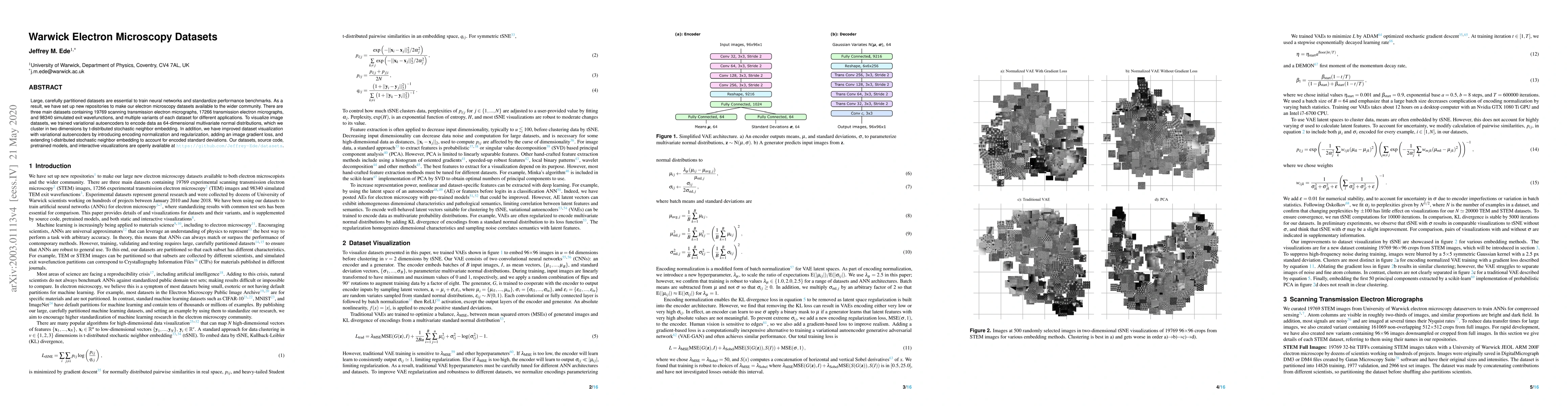
This graph shows how this paper is connected to other research. The current paper is shown in dark gray, papers that cite it are in green, and papers it references are in blue. You can:
• Hover over any node to see the paper title
• Click a node to view that paper's details
• Drag nodes to rearrange the graph
• Use the zoom controls (bottom right) to zoom in/out
Title | Authors | Year | Actions |
---|---|---|---|
Realizing smart scanning transmission electron microscopy using high performance computing.
|
Utkarsh Pratiush, Austin Houston, Sergei V. Kalinin, Gerd Duscher
|
2024
|
View Paper |
Deep Learning Methods for Microstructural Image Analysis: The State-of-the-Art and Future Perspectives
|
Khaled Alrfou, Tian Zhao, Amir Kordijazi
|
2024
|
View Paper |
Low-Resolution-Only Microscopy Super-Resolution Models Generalizing to Non-Periodicities at Atomic Scale
|
Björn Möller, Zhengyang Li, Markus Etzkorn, Tim Fingscheidt
|
2024
|
View Paper |
Probe microscopy is all you need
|
S. Kalinin, R. Vasudevan, Yongtao Liu, Ayana Ghosh, Kevin M. Roccapriore, M. Ziatdinov
|
2023
|
View Paper |
Deep convolutional neural networks to restore single-shot electron microscopy images
|
I. Lobato, T. Friedrich, S. Van Aert
|
2023
|
View Paper |
SIGMA: Spectral Interpretation Using Gaussian Mixtures and Autoencoder
|
Po‐Yen Tung, H. A. Sheikh, M. Ball, F. Nabiei, R. Harrison
|
2023
|
View Paper |
Microscopy is All You Need
|
S. Kalinin, R. Vasudevan, Yongtao Liu, Ayana Ghosh, Kevin M. Roccapriore, M. Ziatdinov
|
2022
|
View Paper |
Computer Vision Methods for the Microstructural Analysis of Materials: The State-of-the-art and Future Perspectives
|
Khaled Alrfou, Amir Kordijazi, Tian Zhao
|
2022
|
View Paper |
Applications of deep learning in electron microscopy.
|
Kevin P. Treder, Chen Huang, Judy S. Kim, A. Kirkland
|
2022
|
View Paper |
Advances in Electron Microscopy with Deep Learning
|
Jeffrey M. Ede
|
2021
|
View Paper |
Deep learning in electron microscopy
|
Jeffrey M. Ede
|
2020
|
View Paper |
Adaptive Partial Scanning Transmission Electron Microscopy with Reinforcement Learning
|
Jeffrey M. Ede
|
2020
|
View Paper |
Exit Wavefunction Reconstruction from Single Transmisson Electron Micrographs with Deep Learning
|
Jeffrey M. Ede, J. J. Peters, J. Sloan, R. Beanland
|
2020
|
View Paper |
Partial Scanning Transmission Electron Microscopy with Deep Learning
|
Jeffrey M. Ede, R. Beanland
|
2019
|
View Paper |
Published: 2021-09-01
Andreas Müller, Martin Weigert, Deborah Schmidt, Lucas Rieckert, Michele Solimena
Published: 2023-03-08
Comments (0)