Centimeter-sized Objects at Micrometer Resolution: Extending Field-of-View in Wavefront Marker X-ray Phase-Contrast Tomography
Publication
Metrics
AI Quick Summary
This paper proposes a novel technique combining eigenflat optimization and deformable image registration to extend the field-of-view in wavefront marker-based X-ray phase-contrast tomography, achieving micrometer resolution for centimeter-sized objects. The method demonstrates successful application on a rat brain sample, overcoming limitations of beam stability and size.
Paper Preview
Abstract
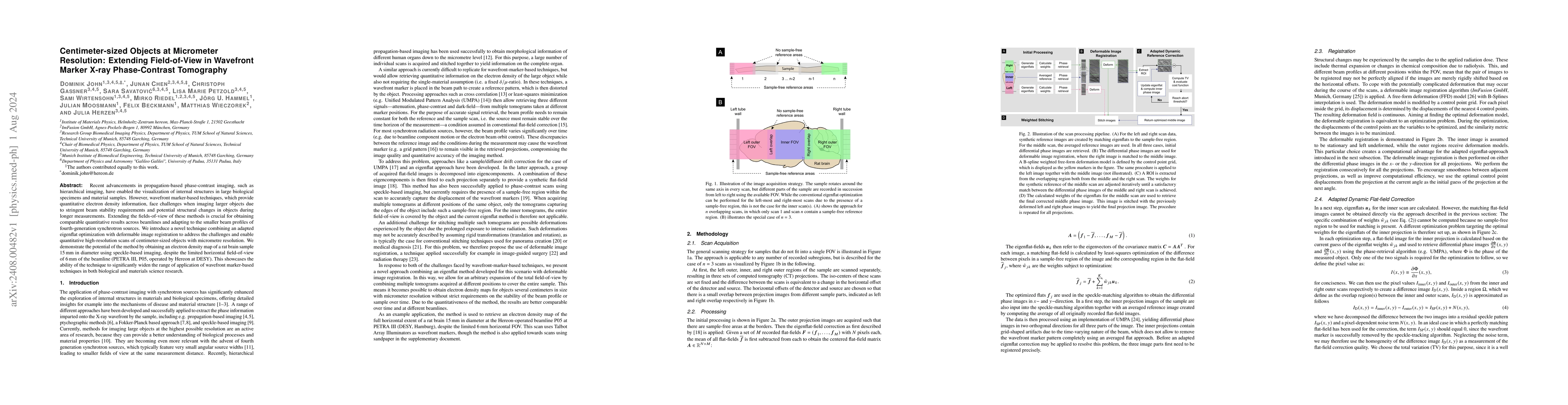
Recent advancements in propagation-based phase-contrast imaging, such as hierarchical imaging, have enabled the visualization of internal structures in large biological specimens and material samples. However, wavefront marker-based techniques, which provide quantitative electron density information, face challenges when imaging larger objects due to stringent beam stability requirements and potential structural changes in objects during longer measurements. Extending the fields-of-view of these methods is crucial for obtaining comparable quantitative results across beamlines and adapting to the smaller beam profiles of fourth-generation synchrotron sources. We introduce a novel technique combining an adapted eigenflat optimization with deformable image registration to address the challenges and enable quantitative high-resolution scans of centimeter-sized objects with micrometre resolution. We demonstrate the potential of the method by obtaining an electron density map of a rat brain sample 15 mm in diameter using speckle-based imaging, despite the limited horizontal field-of-view of 6 mm of the beamline (PETRA III, P05, operated by Hereon at DESY). This showcases the ability of the technique to significantly widen the range of application of wavefront marker-based techniques in both biological and materials science research.
AI Key Findings
Get AI-generated insights about this paper's methodology, results, significance, and more — seven facets brought into focus.
Impact
Paper Details
Authors
PDF Preview
Citation Network
Current paper (gray), citations (green), references (blue)
Display is limited for performance on very large graphs.
Discussion 0