Summary
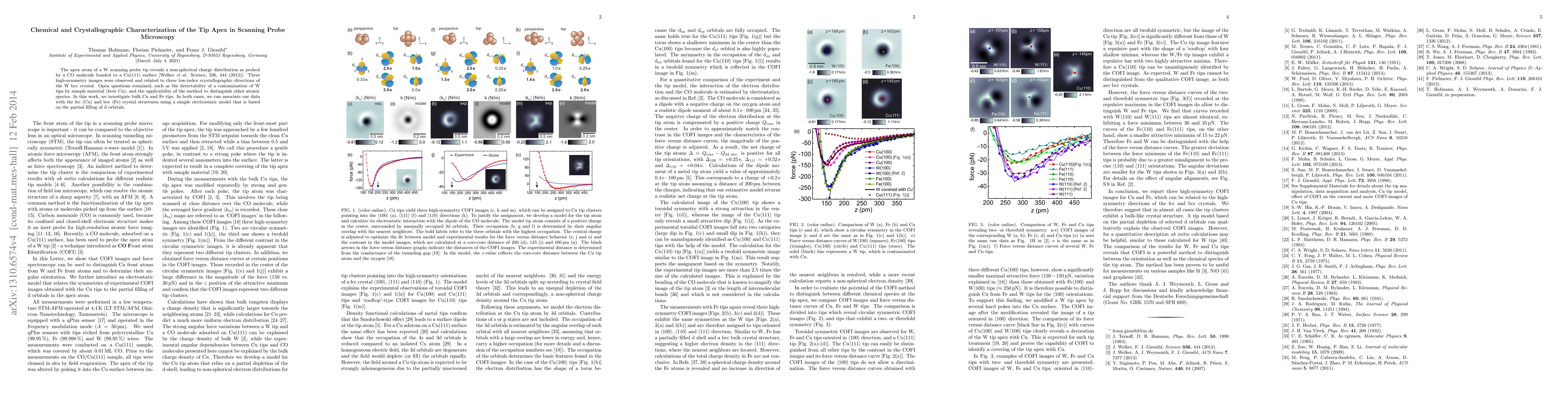
The apex atom of a W scanning probe tip reveals a non-spherical charge distribution as probed by a CO molecule bonded to a Cu(111) surface [Welker et al. Science, 336, 444 (2012)]. Three high-symmetry images were observed and related to three low-index crystallographic directions of the W bcc crystal. Open questions remained, however, including the verification that the tip was indeed W-terminated, and whether this method can be easily applied to distinguish other atomic species. In this work, we investigate bulk Cu and Fe tips. In both cases we can associate our data with the fcc (Cu) and bcc (Fe) crystal structures. A model is presented, based on the partial filling of d orbitals, to relate the AFM images to the angular orientation of the tip structure.
AI Key Findings
Get AI-generated insights about this paper's methodology, results, and significance.
Paper Details
PDF Preview
Key Terms
Citation Network
Current paper (gray), citations (green), references (blue)
Display is limited for performance on very large graphs.
Similar Papers
Found 4 papers| Title | Authors | Year | Actions |
|---|
Comments (0)