Crystallographic image processing for scanning probe microscopy
Publication
Metrics
AI Quick Summary
Researchers have developed a method to process scanning probe microscopy images using crystallography, allowing for correction of distortions and improvement of image quality. This technique uses symmetrized scanning tunneling microscope tips and can be applied to various types of periodic objects.
Paper Preview
Abstract
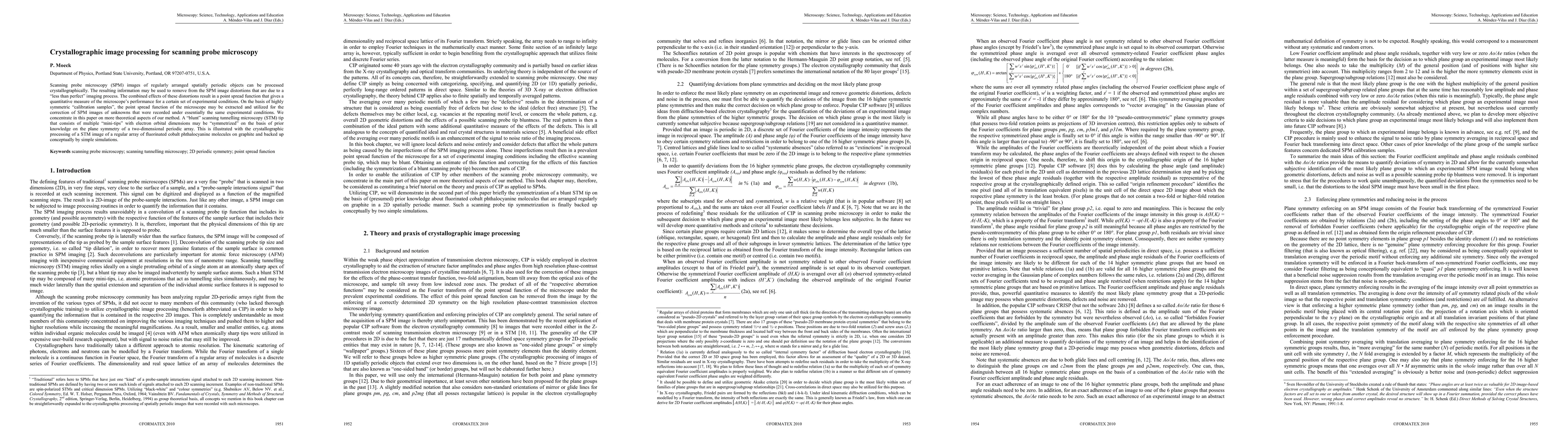
Scanning probe microscopy (SPM) images of regularly arranged spatially periodic objects can be processed crystallographically. The resulting information may be used to remove from the SPM image distortions that are due to a less than perfect imaging process. The combined effects of these distortions result in a point spread function that gives a quantitative measure of the performance of the microscope for a certain set of experimental conditions. On the basis of highly symmetric calibration samples, the point spread function of the microscope may be extracted and utilized for the correction of SPM images of unknowns that were recorded under essentially the same experimental conditions. We concentrate in this paper on more theoretical aspects of our method. A blunt scanning tunneling microscopy (STM) tip that consists of multiple mini-tips with electron orbital dimensions may be symmetrized on the basis of prior knowledge on the plane symmetry of a two-dimensional periodic array. This is illustrated with the crystallographic processing of a STM image of a regular array of fluorinated cobalt phthalocyanine molecules on graphite and backed up conceptually by simple simulations.
AI Key Findings
Get AI-generated insights about this paper's methodology, results, significance, and more — seven facets brought into focus.
Impact
Paper Details
Authors
PDF Preview
Key Terms
Citation Network
Current paper (gray), citations (green), references (blue)
Display is limited for performance on very large graphs.
Discussion 0