Classification of COVID-19 on chest X-Ray images using Deep Learning model with Histogram Equalization and Lungs Segmentation
Publication
Metrics
AI Quick Summary
This paper proposes a deep learning model for classifying COVID-19 on chest X-rays using histogram equalization, U-net segmentation, VGG-16 feature extraction, and SVM classification, achieving a 98% recognition rate. The approach aims to alleviate healthcare pressures by providing an effective diagnostic tool.
Paper Preview
Abstract
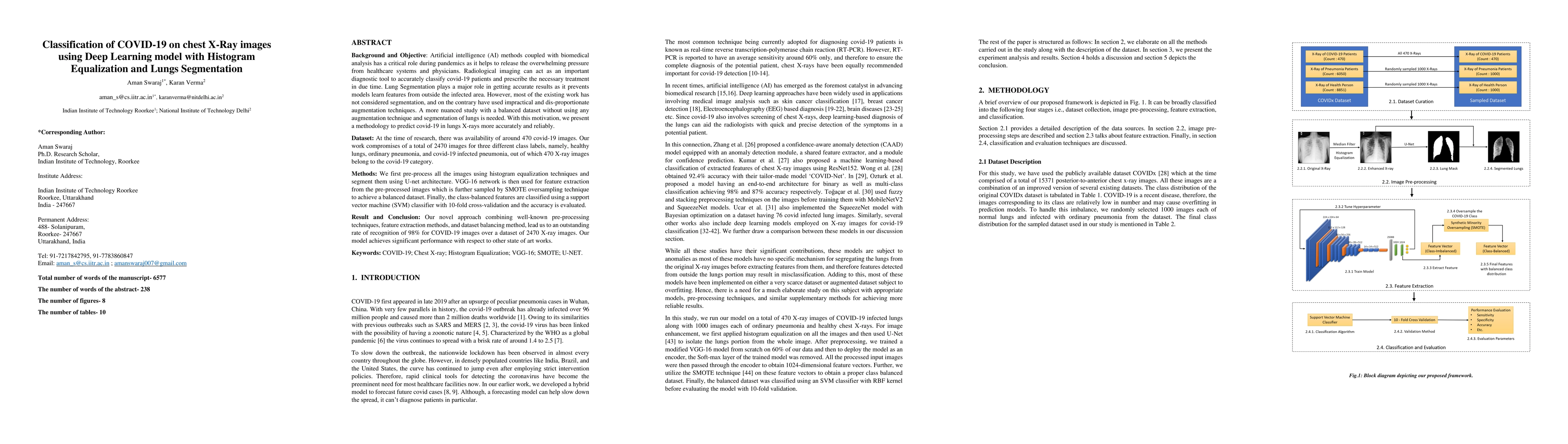
Background and Objective: Artificial intelligence (AI) methods coupled with biomedical analysis has a critical role during pandemics as it helps to release the overwhelming pressure from healthcare systems and physicians. As the ongoing COVID-19 crisis worsens in countries having dense populations and inadequate testing kits like Brazil and India, radiological imaging can act as an important diagnostic tool to accurately classify covid-19 patients and prescribe the necessary treatment in due time. With this motivation, we present our study based on deep learning architecture for detecting covid-19 infected lungs using chest X-rays. Dataset: We collected a total of 2470 images for three different class labels, namely, healthy lungs, ordinary pneumonia, and covid-19 infected pneumonia, out of which 470 X-ray images belong to the covid-19 category. Methods: We first pre-process all the images using histogram equalization techniques and segment them using U-net architecture. VGG-16 network is then used for feature extraction from the pre-processed images which is further sampled by SMOTE oversampling technique to achieve a balanced dataset. Finally, the class-balanced features are classified using a support vector machine (SVM) classifier with 10-fold cross-validation and the accuracy is evaluated. Result and Conclusion: Our novel approach combining well-known pre-processing techniques, feature extraction methods, and dataset balancing method, lead us to an outstanding rate of recognition of 98% for COVID-19 images over a dataset of 2470 X-ray images. Our model is therefore fit to be utilized in healthcare facilities for screening purposes.
AI Key Findings
Get AI-generated insights about this paper's methodology, results, significance, and more — seven facets brought into focus.
Impact
Paper Details
Authors
PDF Preview
Key Terms
Citation Network
Current paper (gray), citations (green), references (blue)
Display is limited for performance on very large graphs.
Discussion 0