Publication
Metrics
AI Quick Summary
This paper compares Generative Adversarial Networks (GANs) with supervised learning methods like CNNs for segmenting Organs at Risk (OAR) in CT scans. The study finds that GAN-based models often perform as well or better than CNN models, especially for complex OAR segmentation tasks.
Paper Preview
Abstract
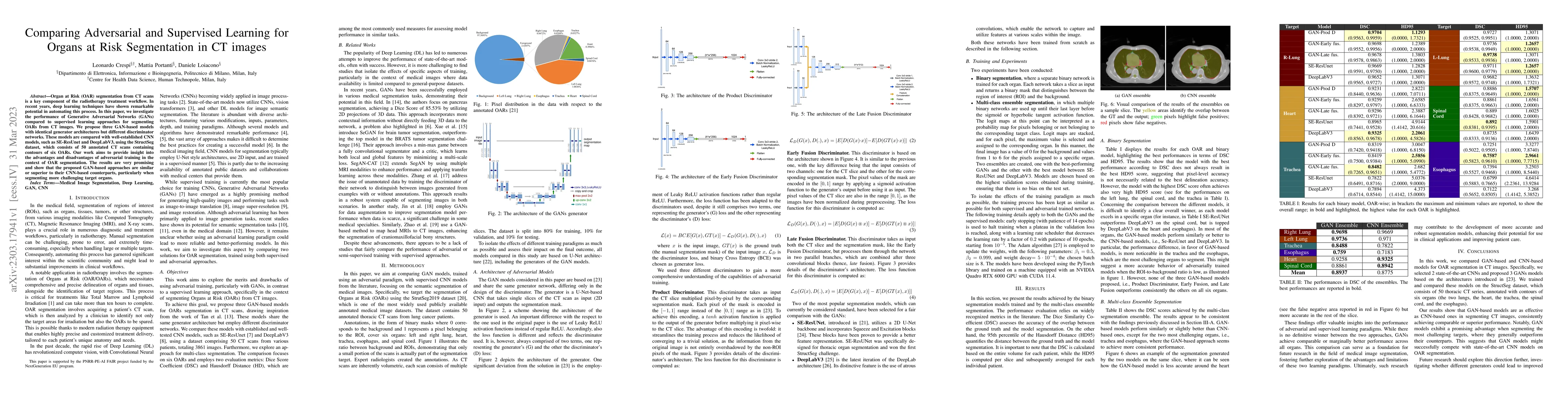
Organ at Risk (OAR) segmentation from CT scans is a key component of the radiotherapy treatment workflow. In recent years, deep learning techniques have shown remarkable potential in automating this process. In this paper, we investigate the performance of Generative Adversarial Networks (GANs) compared to supervised learning approaches for segmenting OARs from CT images. We propose three GAN-based models with identical generator architectures but different discriminator networks. These models are compared with well-established CNN models, such as SE-ResUnet and DeepLabV3, using the StructSeg dataset, which consists of 50 annotated CT scans containing contours of six OARs. Our work aims to provide insight into the advantages and disadvantages of adversarial training in the context of OAR segmentation. The results are very promising and show that the proposed GAN-based approaches are similar or superior to their CNN-based counterparts, particularly when segmenting more challenging target organs.
AI Key Findings
Get AI-generated insights about this paper's methodology, results, significance, and more — seven facets brought into focus.
Impact
Paper Details
Authors
PDF Preview
Key Terms
Citation Network
Current paper (gray), citations (green), references (blue)
Display is limited for performance on very large graphs.
Discussion 0