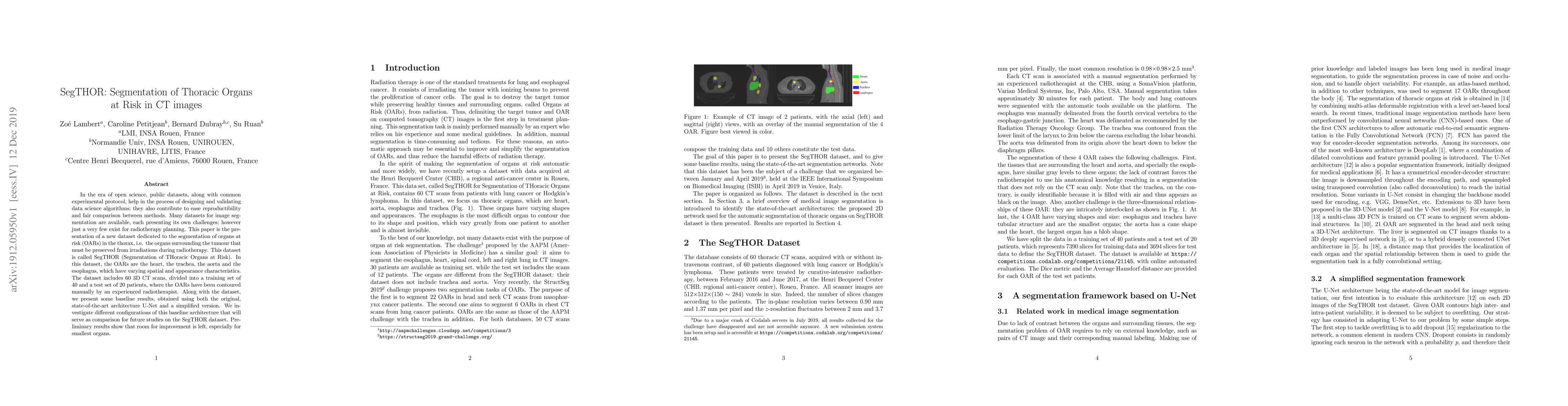
Summary
In the era of open science, public datasets, along with common experimental protocol, help in the process of designing and validating data science algorithms; they also contribute to ease reproductibility and fair comparison between methods. Many datasets for image segmentation are available, each presenting its own challenges; however just a very few exist for radiotherapy planning. This paper is the presentation of a new dataset dedicated to the segmentation of organs at risk (OARs) in the thorax, i.e. the organs surrounding the tumour that must be preserved from irradiations during radiotherapy. This dataset is called SegTHOR (Segmentation of THoracic Organs at Risk). In this dataset, the OARs are the heart, the trachea, the aorta and the esophagus, which have varying spatial and appearance characteristics. The dataset includes 60 3D CT scans, divided into a training set of 40 and a test set of 20 patients, where the OARs have been contoured manually by an experienced radiotherapist. Along with the dataset, we present some baseline results, obtained using both the original, state-of-the-art architecture U-Net and a simplified version. We investigate different configurations of this baseline architecture that will serve as comparison for future studies on the SegTHOR dataset. Preliminary results show that room for improvement is left, especially for smallest organs.
AI Key Findings
Get AI-generated insights about this paper's methodology, results, and significance.
Paper Details
PDF Preview
Key Terms
Citation Network
Current paper (gray), citations (green), references (blue)
Display is limited for performance on very large graphs.
Similar Papers
Found 4 papersComparing Adversarial and Supervised Learning for Organs at Risk Segmentation in CT images
Leonardo Crespi, Daniele Loiacono, Mattia Portanti
| Title | Authors | Year | Actions |
|---|
Comments (0)