This paper proposes a computer method to aid radiologists in detecting microcalcification clusters (MCCs) in mammograms, enhancing images with wavelet transformation and morphology, and comparing adaptive threshold and watershed segmentation methods for MCC segmentation. The goal is to identify the most effective segmentation technique for early breast cancer diagnosis.
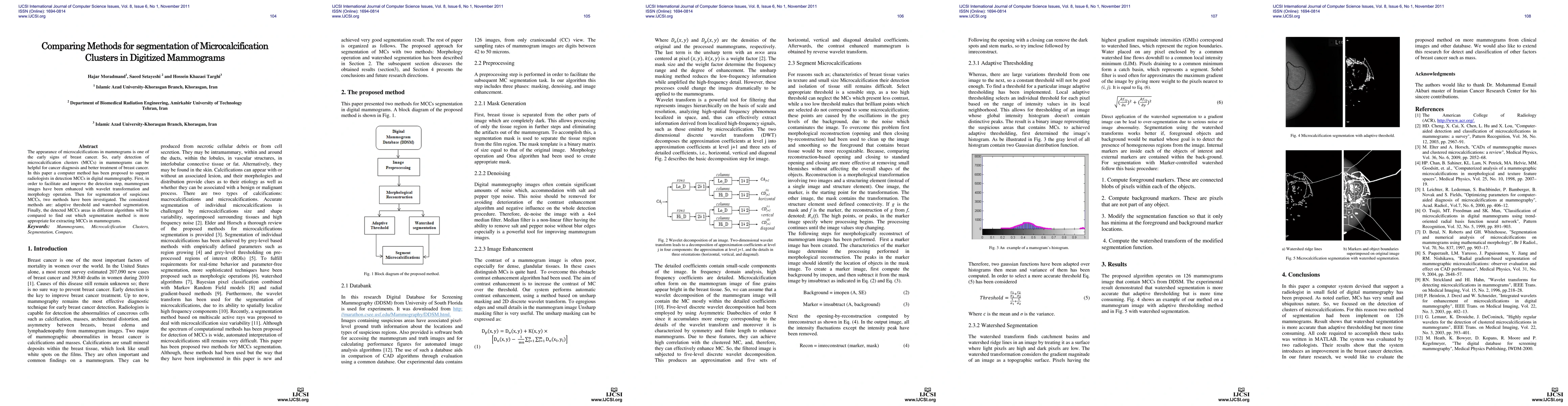
The appearance of microcalcifications in mammograms is one of the early signs of breast cancer. So, early detection of microcalcification clusters (MCCs) in mammograms can be helpful for cancer diagnosis and better treatment of breast cancer. In this paper a computer method has been proposed to support radiologists in detection MCCs in digital mammography. First, in order to facilitate and improve the detection step, mammogram images have been enhanced with wavelet transformation and morphology operation. Then for segmentation of suspicious MCCs, two methods have been investigated. The considered methods are: adaptive threshold and watershed segmentation. Finally, the detected MCCs areas in different algorithms will be compared to find out which segmentation method is more appropriate for extracting MCCs in mammograms.
Get AI-generated insights about this paper's methodology, results, and significance.
Current paper (gray), citations (green), references (blue)
Title | Authors | Year | Actions |
---|---|---|---|
Descriptive analysis of computational methods for automating mammograms with practical applications
|
A. Bhale, M. Joshi
|
2020
|
View Paper |
Classification of Microcalcifications in Mammograms using 2D Discrete Wavelet Transform and Random Forest
|
R. Fadil, A. Jackson, B. A. E. Majd, Hassan El Ghazi, N. Kaabouch
|
2020
|
View Paper |
Intra-Operative Radiotherapy in Breast Cancer Treatment: A Literature Review
|
Alireza Tehrani Yazdi, S. Setayeshi
|
2019
|
View Paper |
Evaluating geodesic active contours in microcalcifications segmentation on mammograms
|
Marcelo A. Duarte, A. Alvarenga, C. Azevedo, M. Calas, A. Infantosi, W. Pereira
|
2015
|
View Paper |
Detection of microcalcification with top-hat transform and the Gibbs random fields
|
Akshay S. Bharadwaj, M. Celenk
|
2015
|
View Paper |
Comparison of Two Segmentation Methods for Mammographic Image
|
Priyanka Jagya, R. Dubey
|
2015
|
View Paper |
Contrast Enhancement of Mammograms for Rapid Detection of Microcalcification Clusters
|
H. Moradmand, S. Setayeshi, A. Karimian, M. Sirous
|
2014
|
View Paper |
An automated method for breast mass segmentation
|
S. Khouaja, H. Jelassi, K. Hamrouni
|
2014
|
View Paper |
Comparing the Performance of Image Enhancement Methods to Detect Microcalcification Clusters in Digital Mammography
|
H. Moradmand, S. Setayeshi, A. Karimian, M. Sirous, M. Akbari
|
2012
|
View Paper |
Segmentation Technique To Detection Microcalcification In Mammograms
|
Ashok Singh Chundawat, Rahul Kumar Malee, Ravita Saraswat, S. Shrivastava
|
2017
|
View Paper |
Classification of Mammographic Masses Using Fuzzy Inference System
|
K. Divyadarshini, R. Vanithamani, S. Sharmili
|
2015
|
View Paper |
Improved Segmentation of Suspicious Regions of Masses in Mammograms by Watershed Transform
|
P. Anuradha, B. R. Jose, J. Mathew
|
2015
|
View Paper |
Segmenting mammographic microcalcifi cations using a semi-automatic procedure based on Otsu ’ s method and morphological fi lters
|
Marcelo A. Duarte, A. Alvarenga, C. Azevedo, M. J. Gregório, Calas, A. Infantosi, W. Pereira
|
2013
|
View Paper |
Study on Microcalcification Detection Using Fisher Discriminant and SVM
|
Jinghuan Guo, Shenglai Chen, Ge Ku, Zhaoqian Sun
|
2013
|
View Paper |
An investigation of the breast cancer classification using various machine learning techniques
|
R. Tripathy
|
2013
|
View Paper |
Handbook of X-ray Imaging Physics and Technology
|
B. Illerhaus, Andreas Staude
|
View Paper |
Comments (0)