ArxivLens

Quick Summary:

This paper introduces a new method for detecting microcalcification clusters in mammograms using Fractal Dimension and Hurst co-efficient, outperforming the conventional Sobel method by replacing its Fudge factor with an image-dependent Hurst co-efficient. The results demonstrate superior edge detection accuracy.

arXiv Id: 1304.8092
Comments: Appeared in ICECIT-2012
Date Published: 2013-05-01
Date Updated: 2013-05-01

Categories:

Key Terms (TF-IDF):

fractal (0.282)
image (0.262)
image (0.252)
efficient (0.248)
dimension (0.247)
efficient (0.237)
technique (0.237)
edges (0.233)
factor (0.231)
technique (0.230)
dimension (0.225)
edges (0.224)
factor (0.222)
method (0.209)
method (0.200)
images (0.193)
dependent (0.188)
images (0.183)
dependent (0.174)
proposed (0.171)
License: http://arxiv.org/licenses/nonexclusive-distrib/1.0/

Summary:

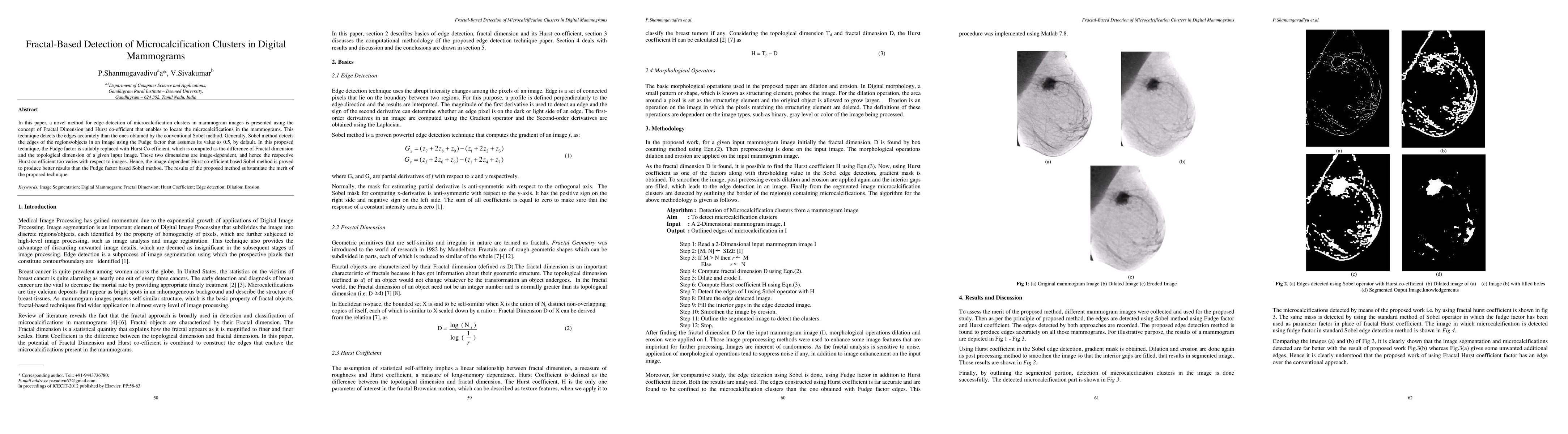
In this paper, a novel method for edge detection of microcalcification clusters in mammogram images is presented using the concept of Fractal Dimension and Hurst co-efficient that enables to locate the microcalcifications in the mammograms. This technique detects the edges accurately than the ones obtained by the conventional Sobel method. Generally, Sobel method detects the edges of the regions/objects in an image using the Fudge factor that assumes its value as 0.5, by default. In this proposed technique, the Fudge factor is suitably replaced with Hurst Co-efficient, which is computed as the difference of Fractal dimension and the topological dimension of a given input image. These two dimensions are image-dependent, and hence the respective Hurst co-efficient too varies with respect to images. Hence, the image-dependent Hurst co-efficient based Sobel method is proved to produce better results than the Fudge factor based Sobel method. The results of the proposed method substantiate the merit of the proposed technique.

Citation & Reference Network Visualization

This graph shows how this paper is connected to other research. The current paper is shown in dark gray, papers that cite it are in green, and papers it references are in blue. You can:

Current Paper
Papers citing this work (Citations)
Papers cited by this work (References)

• Hover over any node to see the paper title

• Click a node to view that paper's details

• Drag nodes to rearrange the graph

• Use the zoom controls (bottom right) to zoom in/out

Citations and References

Title Authors Year Actions
Analysis of Mammography Techniques for Breast Cancer Detection
P. K. Diderot, N. Vasudevan, V. R. Prakash
2019
View Paper
Detection of Breast Cancer in Mammogram Images Using Texture Analysis Methods
A. M. Omer, A. O. H. Supervisor
2019
View Paper
The automatic detection of hiding narcotics in human bodies based on fractal dimension and SVM classification
Li Weijiao, Chen Jiamin, Wu Xiaomei, Wang Weiqi
2017
View Paper
Fuzzy Based Contrast Enhancement Method for Lung Cancer CT Images
R. Khare
2017
View Paper
A Survey on the Analysis of Segmentation Techniques in Mammogram Images
M. P. Sukassini, T. Velmurugan
2015
View Paper
Spectral correlation analysis for microcalcification detection in digital mammogram images
Aya F. Khalaf, I. Yassine
2015
View Paper
WAVELET TRANSFORMATION-BASED DETECTION OF MASSES IN DIGITAL MAMMOGRAMS
P. Shanmugavadivu, V. Sivakumar, J. Suhanya
2014
View Paper
Analysis of Segmentation Methods for Locating Microcalcification in Mammogram Image
J. Jebathangam, S. Purushothaman
2016
View Paper
A Novel Technique for Mammogram Mass Segmentation Using Fractal Adaptive Thresholding
P. Shanmugavadivu, V. Sivakumar
2014
View Paper

Comments (0)

This website uses cookies to ensure you get the best experience. Learn more