Summary
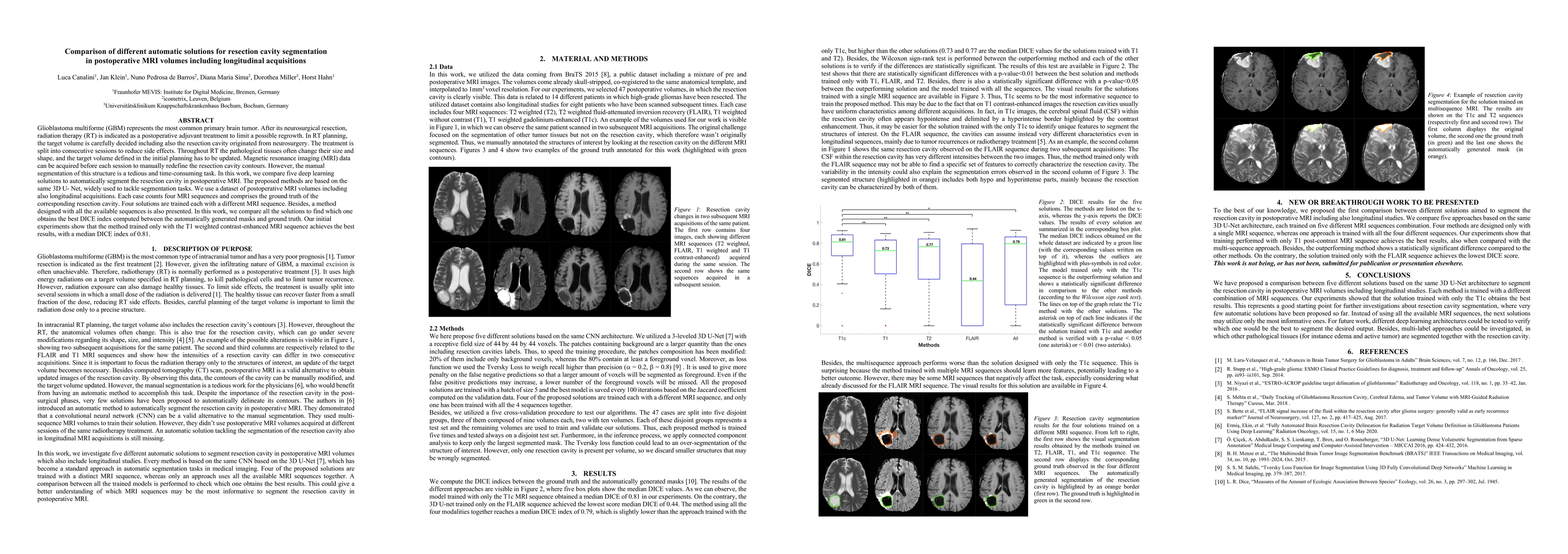
In this work, we compare five deep learning solutions to automatically segment the resection cavity in postoperative MRI. The proposed methods are based on the same 3D U-Net architecture. We use a dataset of postoperative MRI volumes, each including four MRI sequences and the ground truth of the corresponding resection cavity. Four solutions are trained with a different MRI sequence. Besides, a method designed with all the available sequences is also presented. Our experiments show that the method trained only with the T1 weighted contrast-enhanced MRI sequence achieves the best results, with a median DICE index of 0.81.
AI Key Findings
Generated Sep 05, 2025
Methodology
A comparison between five different solutions based on the same 3D U-Net architecture to segment the resection cavity in postoperative MRI volumes including longitudinal studies.
Key Results
- The solution trained with only the T1c sequence achieves the best results.
- The multisequence approach performs worse than the single-sequence approach.
- The method trained on FLAIR sequence achieves the lowest DICE score.
- The 3D U-Net architecture is effective for segmenting resection cavities in postoperative MRI volumes.
Significance
This research contributes to the development of automatic solutions for segmenting resection cavities in postoperative MRI volumes, which is crucial for radiation therapy planning and patient treatment.
Technical Contribution
The proposed comparison between different solutions based on the same 3D U-Net architecture provides a novel approach for evaluating segmentation performance and identifying the most effective architectures for resection cavity segmentation.
Novelty
This work is novel because it compares multiple approaches using the same architecture, providing insights into the effectiveness of different MRI sequences for segmenting resection cavities.
Limitations
- The dataset used in this study may not be representative of all cases.
- The 3D U-Net architecture may not generalize well to other segmentation tasks.
Future Work
- Investigating the use of transfer learning for segmenting resection cavities in postoperative MRI volumes.
- Developing multi-label approaches for segmenting multiple pathological tissues simultaneously.
Paper Details
PDF Preview
Key Terms
Citation Network
Current paper (gray), citations (green), references (blue)
Display is limited for performance on very large graphs.
Similar Papers
Found 4 papersRefining cardiac segmentation from MRI volumes with CT labels for fine anatomy of the ascending aorta.
Oda, Hirohisa, Wakamori, Mayu, Akita, Toshiaki
| Title | Authors | Year | Actions |
|---|

Comments (0)