Publication
Metrics
AI Quick Summary
This paper proposes a convolutional neural network (CNN) method to automatically characterize plaques in intracoronary optical coherence tomography (OCT) images, aiming to improve the efficiency and accuracy of plaque analysis compared to traditional machine learning methods. The CNN approach demonstrated an average prediction accuracy of 0.866 on 269 OCT images, showing potential for clinical use.
Paper Preview
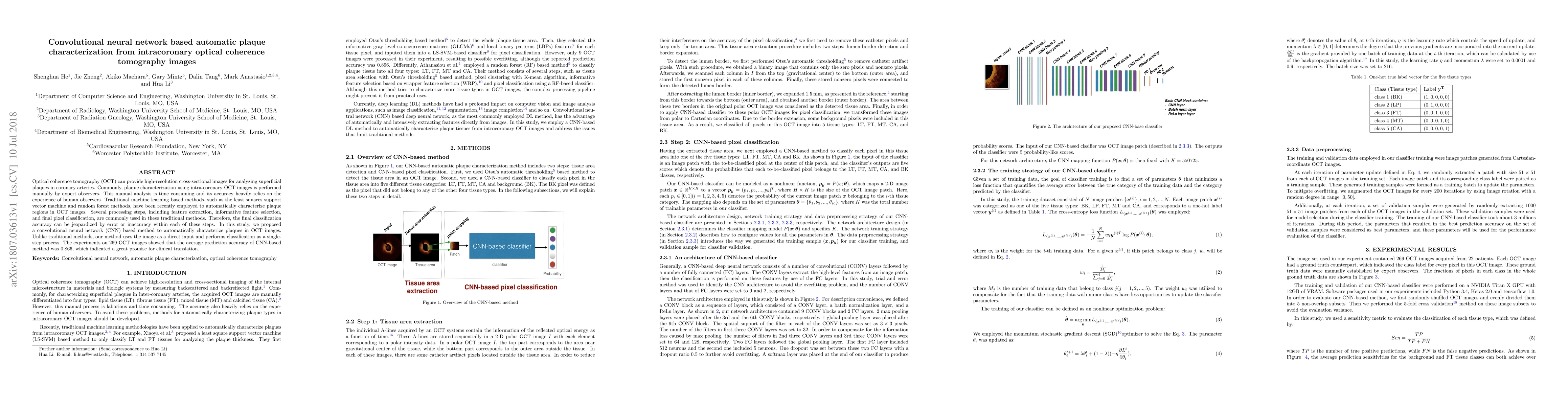
Abstract
Optical coherence tomography (OCT) can provide high-resolution cross-sectional images for analyzing superficial plaques in coronary arteries. Commonly, plaque characterization using intra-coronary OCT images is performed manually by expert observers. This manual analysis is time consuming and its accuracy heavily relies on the experience of human observers. Traditional machine learning based methods, such as the least squares support vector machine and random forest methods, have been recently employed to automatically characterize plaque regions in OCT images. Several processing steps, including feature extraction, informative feature selection, and final pixel classification, are commonly used in these traditional methods. Therefore, the final classification accuracy can be jeopardized by error or inaccuracy within each of these steps. In this study, we proposed a convolutional neural network (CNN) based method to automatically characterize plaques in OCT images. Unlike traditional methods, our method uses the image as a direct input and performs classification as a single-step process. The experiments on 269 OCT images showed that the average prediction accuracy of CNN-based method was 0.866, which indicated a great promise for clinical translation.
AI Key Findings
Get AI-generated insights about this paper's methodology, results, significance, and more — seven facets brought into focus.
Impact
Paper Details
PDF Preview
Key Terms
Citation Network
Current paper (gray), citations (green), references (blue)
Display is limited for performance on very large graphs.
Discussion 0